デジタル顕微鏡

デジタル顕微鏡

デジタル顕微鏡

デジタル顕微鏡は、接眼レンズのない顕微鏡です。 デジタルカメラが内蔵されています。 ライブ像がモニター画面に表示され、複数人で共有、画像保存、レポート出力に適しています。

接眼レンズ不要の検査、記録、デジタル解析

ライカのデジタル顕微鏡は高い光学性能と使いやすい操作で、記録、解析、生産、品質管理、品質保証、故障解析などに最適です。

170年以上の光学顕微鏡の歴史とデジタルとの融合でお客様ニーズにこたえます。

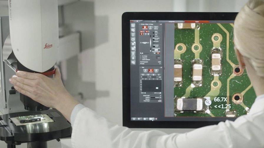
ライカのデジタル顕微鏡が選ばれる理由とは?

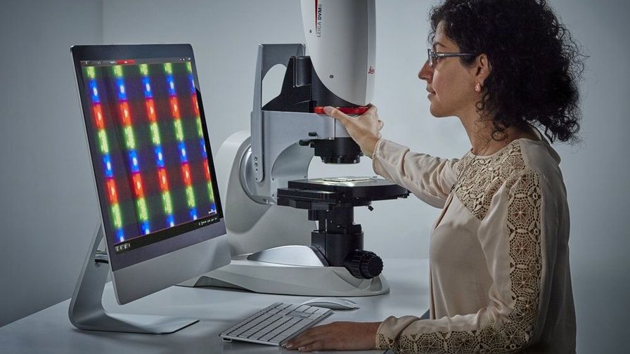
高い光学性能で高い色再現性と、使いやすいソフトウェアでサンプルに忠実なデータを引き出します。

簡単な操作

わかりやすいアイコンで直感的に操作できます。 例: 片手操作でレンズを交換する方法を紹介しています。
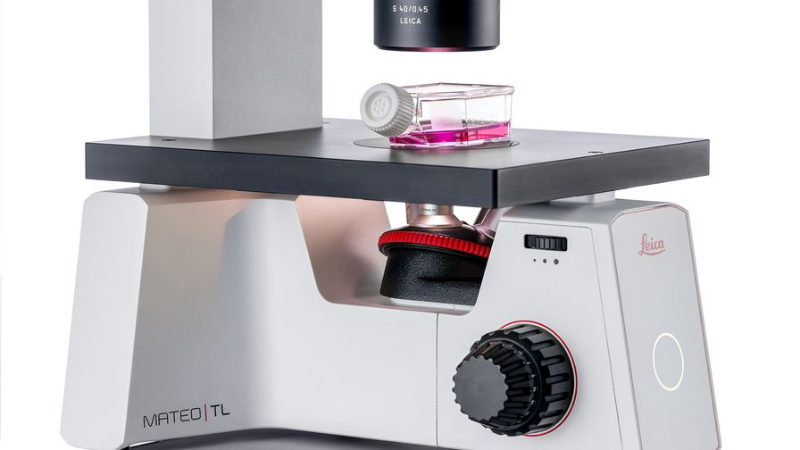
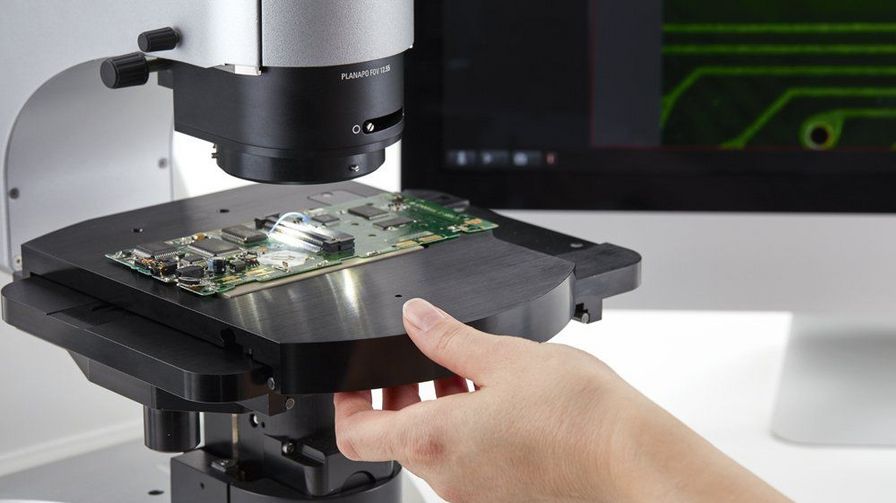
スムーズなワークフロー

観察からレポートの作成、分析まで、ワークフローベースで進めることができます。 さらに、ソフトウェアが処理をスピードアップさせます。

綺麗な画像

ライカの高い光学はその優れた技術で世界で評価されています。 高い光学性能と最新カメラで、サンプルのあらゆる細部を捉えた、色再現性に優れた画像を生成します。

快適な姿勢

ライカのデジタル顕微鏡は、顕微鏡ワークステーションをコンピュータワークプレイスに変えます。 まっすぐに自然な姿勢で操作できるようにデザインされています。

高い柔軟性

大きなサンプルにも小さなサンプルにも対応できます。 焦点深度が深く、何度もピント合わせたりする必要がありません。

安心

取得条件の自動保存で高い再現性を提供します。 顕微鏡の設定やカメラ条件の読み取り機能が、これを可能にしています。

簡単操作と高い信頼性ソフトウェア

デジタル顕微鏡は、目視しないためソフトウェアがキーです。 複雑な画像解析機能も、簡単操作でサポートします。

主な機能

  • 画像および焦点合成画像の取得
  • 2Dと3D測定
  • カメラ、顕微鏡条件など画像取得時に同時保存
  • レポートの作成

傾斜スタンド

傾斜スタンドが一体化されており、片手操作でさまざまな角度からサンプルを観察できます。 これは特に次の検査に役立ちます。

  • 金属パーツの腐食パターンなどの複雑な構造
  • 故障解析における欠陥部位の検出
  • 半導体生産に使用されるボンド
  • 異物の1種である昆虫の観察

サンプルの位置はそのままで、 スタンド傾斜またはステージ回転するだけで、サンプルのさまざまな状況を観察でき、新たな発見につながります。

簡単操作で、作業に集中

デジタル顕微鏡には他にもメリットがあります。

経験の少ないビギナーの方でも、作業に短時間で慣れることができ、高解像、きれいな画像を取得して、画面で直接品質をチェックできます。
 

関連記事

デジタル顕微鏡に関する最新記事を読む

ライカ マイクロシステムズのサイエンスラボでは、 顕微鏡に関する専門記事やお客様事例、基本情報を提供しています。 日常業務や研究で、ビギナーから経験豊富な方まで幅広くサポートします。

その他の記事
Example of calibrating a microscope at a higher magnification value using a stage micrometer.

Microscope Calibration for Measurements: Why and How You Should Do It

Microscope calibration ensures accurate and consistent measurements for inspection, quality control (QC), failure analysis, and research and development (R&D). Calibration steps are described in this…
Mouse brain slice which was immunostained with GFAP-A647 and imaged using a THUNDER Imager Tissue. Courtesy of H. Xu, University of Pennsylvania, Philadelphia, USA.

神経科学研究

神経変性疾患の理解向上に取り組んでいる、もしくは神経系の機能を研究をしていますか? ライカマイクロシステムズのイメージングソリューションによってブレイクスルーを起こす方法をご覧ください。
Evolved ARveo and MyVeo in Operating Room

The Guide to Augmented Reality in Microsurgery

In an era of technological advancement, Augmented Reality (AR) is rapidly transforming the medical field. In surgical microscopy, AR can display fluorescence signals as digital overlays in real-time…
Documentation of an automotive clutch friction surface with a digital microscope

Automotive Part Verification and Development according to Specifications

Automotive part verification during the development and production of parts and components by suppliers or manufacturers is important for ensuring that specifications are met. Specifications are…
3D culture of ovarian cancer cells imaged using the confocal mode of Mica.

Mica: A Game-changer for Collaborative Research at Imperial College London

This interview highlights the transformative impact of Mica at Imperial College London. Scientists explain how Mica has been a game-changer, expanding research possibilities and facilitating…
Area of a printed circuit board (PCB) which was imaged with extended depth of field (EDOF) using digital microscopy.

顕微鏡を知る:被写界深度

顕微鏡において被写界深度は、凹凸の変化が⼤きい構造を持つ試料をピントがあったシャープに観察・撮像するために重要なパラメータです。被写界深度は、開⼝数、解像度、倍率の相関関係によって決定され、解像度とパラメータは反⽐例の関係にあります。被写界深度と解像度のバランスが最適になるように調整することができる顕微鏡もあります。
Digital microscopy simplifies documenting cell-culture results electronically while following 21 CFR part 11 guidelines for biopharma.

Introduction to 21 CFR Part 11 for Electronic Records of Cell Culture

This article provides an introduction to the recommendations of 21 CFR Part 11 from the FDA, specifically focusing on the audit trail and user management in the context of cell-culture laboratories.…
Blood vessel system of a zebrafish larvae

Overcoming Challenges with Microscopy when Imaging Moving Zebrafish Larvae

Zebrafish is a valuable model organism with many beneficial traits. However, imaging a full organism poses challenges as it is not stationary. Here, this case study shows how zebrafish larvae can be…
GLOW800 Augmented Reality Fluorescence used for real-time blood flow visualization in aneurysm clipping surgery

Aneurysm Clipping: Assessing Perforators in Real-Time with AR Fluorescence

This article covers two aneurysm clipping cases highlighting the clinical benefits of GLOW800 Augmented Reality Fluorescence application in neurosurgery, based on insights from Prof. Tohru Mizutani,…
GLOW400 AR Fluorescence in anatomy view, image courtesy of Prof. Kondo

How to Achieve Brain Tissue Resection with GLOW400 AR

Intraoperative MRI is one form of real-time intraoperative visualization, but if more in-depth visualization to identify a tumor during surgery is wanted, intraoperative fluorescence diagnostics is…
Salmonella biofilms 3D render

Exploring Microbial Worlds: Spatial Interactions in 3D Food Matrices

The Micalis Institute is a joint research unit in collaboration with INRAE, AgroParisTech, and Université Paris-Saclay. Its mission is to develop innovative research in the field of food microbiology…
Immunofluorescence image of a mouse enodmetrial organoid stained with CK14 and DAPI

Advancing Uterine Regenerative Therapies with Endometrial Organoids

Prof. Kang's group investigates important factors that determine the uterine microenvironment in which embryo insertion and pregnancy are successfully maintained. They are working to develop new…
Dapi – Nucleus, GFP – Plasma Membrane, Thickness 100µm, 63x objektive, 469 Z planes, 2 channels, THUNDER Imager 3D Cell Culture. Courtesy M.Sc. Dana Krauß, Medical University of Vienna (Austria).

How Efficient is your 3D Organoid Imaging and Analysis Workflow?

Organoid models have transformed life science research but optimizing image analysis protocols remains a key challenge. This webinar explores a streamlined workflow for organoid research, starting…
AI-based transfection analysis (left) of U2OS cells which were transfected with a fluorescently labelled protein. A fluorescence image of the cells (right) is also shown. The analysis and imaging were performed with Mateo FL.

Leveraging AI for Efficient Analysis of Cell Transfection

This article explores the pivotal role of artificial intelligence (AI) in optimizing transfection efficiency measurements within the context of 2D cell culture studies. Precise and reliable…
AI-based cell counting performed with a phase-contrast and fluorescence image using the Mateo FL microscope.

Precision and Efficiency with AI-Enhanced Cell Counting

This article describes the use of artificial intelligence (AI) for precise and efficient cell counting. Accurate cell counting is important for research with 2D cell cultures, e.g., cellular dynamics,…
Image of confluent cells taken with phase contrast (left) and analyzed for confluency using AI (right).

AI Confluency Analysis for Enhanced Precision in 2D Cell Culture

This article explains how efficient, precise confluency assessment of 2D cell culture can be done with artificial intelligence (AI). Assessing confluency, the percentage of surface area covered,…
Evolved ARveo 8: Operating Room (OR) set-up.

Augmented Reality: Transforming Neurosurgical Procedures

In this ebook, you will explore the exciting advances that Augmented Reality (AR) brings to the field of neurosurgery. This comprehensive guide, including explanatory videos, addresses key questions…
The role of extracellular signalling mechanisms in the correct development of the human brain

How do Cells Talk to Each Other During Neurodevelopment?

Professor Silvia Capello presents her group’s research on cellular crosstalk in neurodevelopmental disorders, using models such as cerebral organoids and assembloids.
Mateo TL inside laminar flow hood user

細胞培養

ライカが提供する培養細胞・培養組織用倒立顕微鏡は、ユーザーが必要とする機能を装備し、ライブセルイメージングのワークフローの効率を向上させます。

もっと読む

神経学

脳神経外科用顕微鏡

ライカ マイクロシステムズは、最も複雑なマイクロサージャリー アプリケーションで脳神経外科医をサポートするための各種外科用顕微鏡ソリューションを提供しています。

もっと読む

Medical Device Manufacturing

医療機器製造

医療機器の生産と開発には、デジタル顕微鏡が欠かせません。 高いレギュレーションが要求される中、欠陥部位などをすぐに特定、検証し、記録必要です。

もっと読む

Automotive and Transportation Sample

自動車と輸送機械

EV(電気自動車)の動力源がエンジンからモーターとなることで、モーターそのものや、バッテリーなど、EVに不可欠な部品の需要が急速に拡大しています。 大容量バッテリーを搭載した自動車の増加に伴い、発熱・発火の原因となる部品・部材の金属コンタミへの注意も重要度が増しています。 ISO、VDAなどの国際規格、地域規格が厳格さを増す中、清浄度評価はコンプライアンスの達成に必須となっています。 ライカのコンタミネーション(残留異物)解析システムは、 粒子のサイズと個数、形状と分類を簡単に解析します。

もっと読む

Mateo TL inside laminar flow hood user
神経学
Medical Device Manufacturing
Automotive and Transportation Sample
Scroll to top