Session has expired. If you were logged in then please login again.
提案
ドロップダウンメニューから国を選択して、位置に関連する追加のパーソナライズされたコンテンツを受け取ってください。デフォルトでは、接続元の国が既に選択されています。
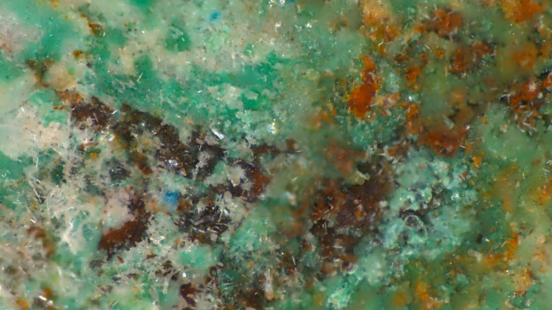
ライカマイクロシステムズの電顕用試料作製装置は、医学・生物学用、産業用共に幅広い製品群で、様々なニーズにお応えします。
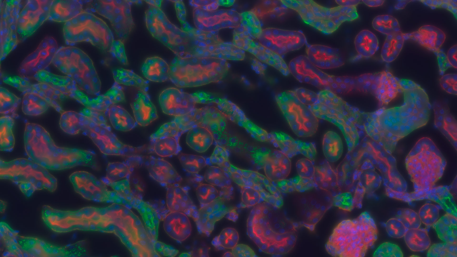
ライカマイクロシステムズのライフサイエンスリサーチ部門は、革新的技術と理化学分野の深い専門知を要する業界において、微細構造の可視化、測定、分析のためのイメージングソリューションを提供します。
もっと読む
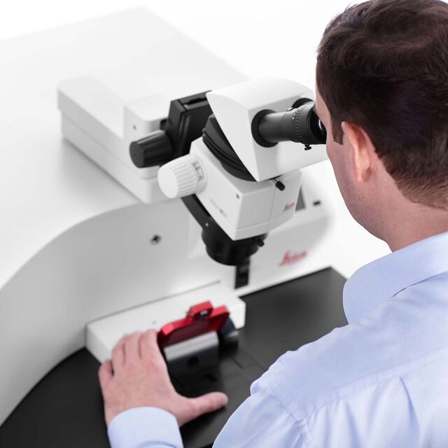
信頼性が高く、高品質なイメージングと解析を行なうためには、適切なツールが必要です。顕微鏡のライカは、 幅広い光学機器、イメージングシステム、ソフトウェア、人間工学に基づいたアクセサリで、お客様のニーズに合った最適なソリューションを提供致します。
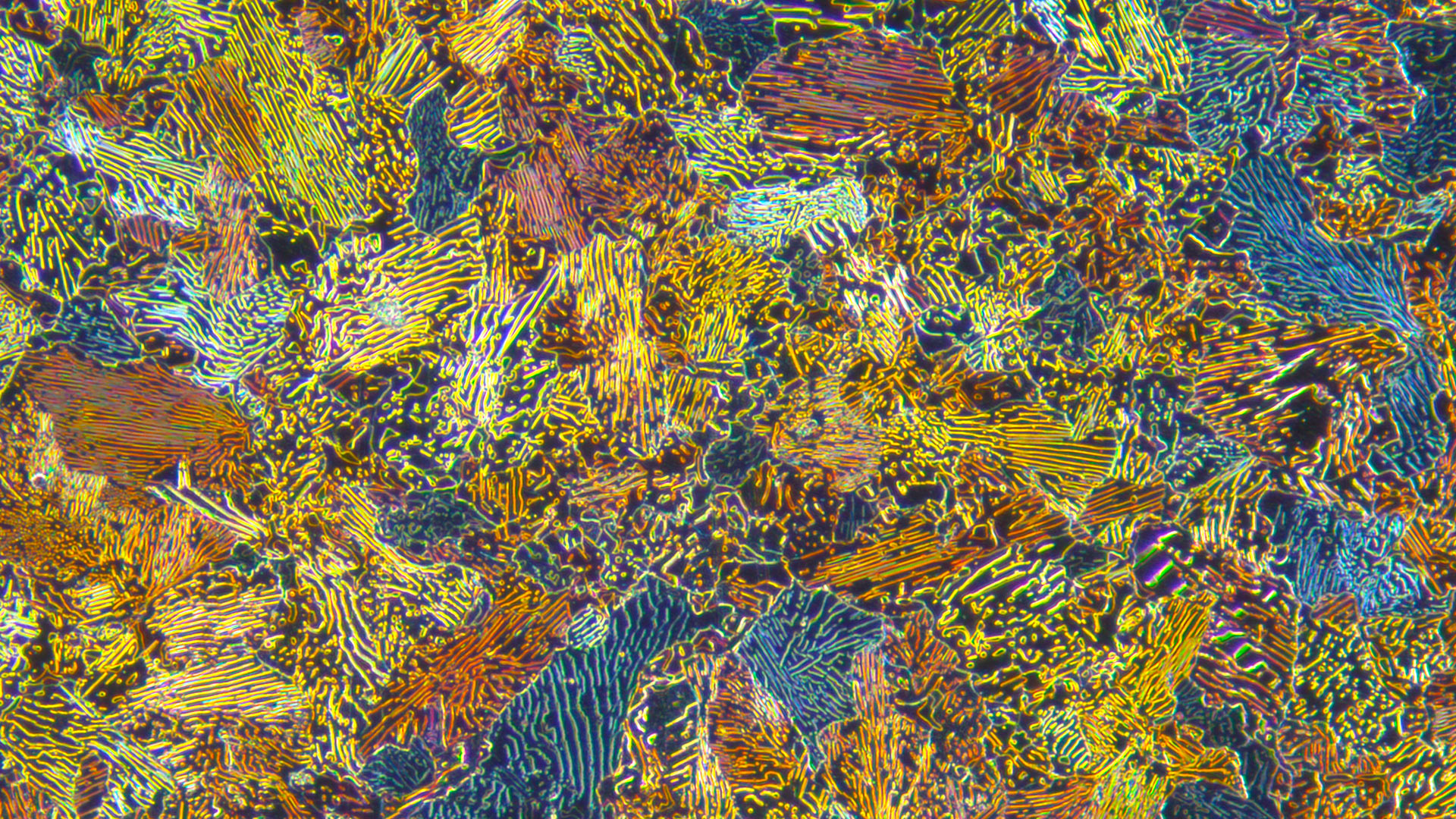
顕微鏡のライカマイクロシステムズは、無駄の軽減、仕事のクオリティ向上に役立つ製品の開発に取り組んでいます。生産活動におけるさまざまな対象物を正確に認識・理解し、効率的にレポート作成していただけるよう、幅広い製品ラインナップの中から、 お客様のワークフローに最適な製品をご提案致します。
教師と生徒、みんなが、課題に集中できると、顕微鏡観察の授業は格段に楽しくなります。ライカの顕微鏡は、 実践的で扱いやすく、生徒をくぎづけにする顕微鏡画像が子どもたちの可能性を引き出します。
サービス内容および見積
現地またはリモートでのデモ
I need help keeping my system running: technical service, repairs, spare parts, upgrades or software licenses.
システムの操作のサポートおよびトレーニング
ライカまでお気軽にご相談ください Show local contacts
フォームを閉じてよろしいですか?
no yes