Industriel
Industriel
Plongez dans des articles détaillés et des webinaires consacrés à l'inspection efficace, à l'optimisation des flux de travail et au confort ergonomique dans les contextes industriels et pathologiques. Les sujets abordés comprennent le contrôle de la qualité, l'analyse des matériaux, la microscopie en pathologie, parmi beaucoup d'autres. C'est ici que vous obtiendrez des informations précieuses sur l'utilisation des technologies de pointe pour améliorer la précision et l'efficacité des processus de fabrication, ainsi que pour établir des diagnostics et des recherches pathologiques précis.
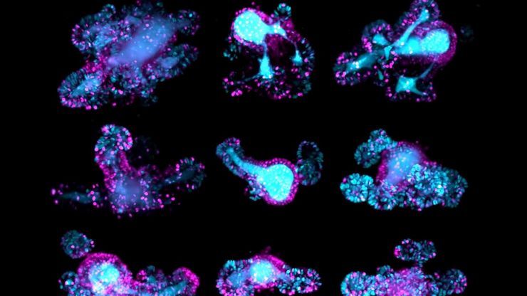
Focus on Long-Term Imaging in 3D with Light Sheet Microscopy
Long-term 3D imaging reveals how complex multicellular systems grow and develop and how cells move and interact over time, unlocking critical insights into development, disease, and regeneration.…
Capturing Developmental Dynamics in 3D
This application note showcases how the Viventis Deep dual-view light sheet microscope was successfully used by researchers for exploring high-resolution, long-term imaging of 3D multicellular models…
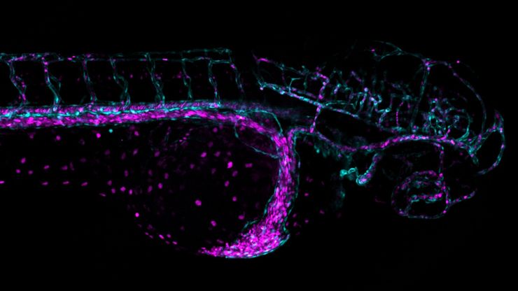
A Guide to Zebrafish Research
To obtain optimal results while doing zebrafish research, especially during screening, sorting, handling, and imaging, seeing the fine details and structures is important. They help researchers make…
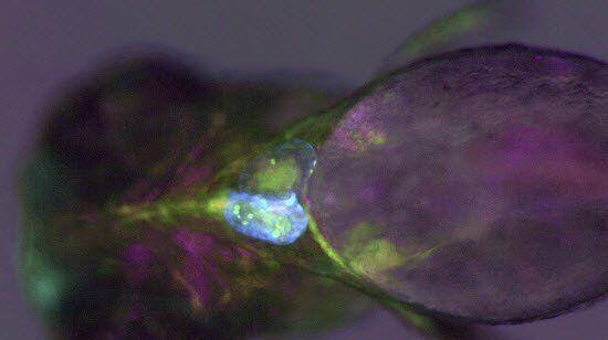
Improving Zebrafish-Embryo Screening with Fast, High-Contrast Imaging
Discover from this article how screening of transgenic zebrafish embryos is boosted with high-speed, high-contrast imaging using the DM6 B microscope, ensuring accurate targeting for developmental…
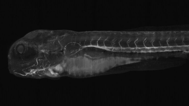
Overcoming Challenges with Microscopy when Imaging Moving Zebrafish Larvae
Zebrafish is a valuable model organism with many beneficial traits. However, imaging a full organism poses challenges as it is not stationary. Here, this case study shows how zebrafish larvae can be…
Using U-Shaped Glass Capillaries for Sample Mounting
The DLS microscope system from Leica Microsystems is an innovative concept which integrates the Light Sheet Microscopy technology into the confocal platform. Due to its unique optical architecture,…
Imaging and Analyzing Zebrafish, Medaka, and Xenopus
Discover how to image and analyze zebrafish, medaka, and Xenopus frog model organisms efficiently with a microscope for developmental biology applications from this article.


