Gold EM grid with a silicon dioxide (SiO₂) support film, containing vitrified T47D cells intrinsically expressing GFP-H2B (labeling nuclei) and RFP-LifeAct (labeling the actin cytoskeleton) and high-pressure frozen C. elegans germline tissue, fluorescently labeled with GFP-tagged synaptonemal complex protein

Flussi di lavoro per la preparazione dei campioni EM e i loro Usi

I ricercatori possono ottenere risultati di alta qualità, precisi e riproducibili durante l'acquisizione di campioni con la Microscopia elettronica utilizzando le soluzioni Leica per la preparazione dei campioni. Le nostre soluzioni supportano la microscopia elettronica a scansione (SEM), la Microscopia elettronica a trasmissione (TEM) e la crio EM.

Contattaci!

I nostri esperti di imaging sono a disposizione per fornire consigli sulle soluzioni per i flussi di lavoro di preparazione dei campioni EM.

Come possono i ricercatori preparare i campioni per il SEM?

La preparazione dei campioni per la SEM (Microscopia elettronica a scansione) prevede tecniche quali il rivestimento, l'essiccazione e l'inclusione per garantire un'immagine ottimale. Utilizzando le soluzioni Leica per i campioni SEM, i ricercatori migliorano la conduttività, riducono gli artefatti e garantiscono superfici stabili e di alta qualità per un'imaging SEM affidabile a temperatura ambiente.

Quali sono i metodi migliori per la preparazione dei campioni TEM?

La preparazione dei campioni TEM (microscopia elettronica a trasmissione) richiede sezioni ultrasottili e prive di danni, rivestimento sputter di griglie EM e colorazione per ottenere una visualizzazione dettagliata della struttura. Con la precisione e l'affidabilità degli strumenti Leica, i ricercatori possono produrre immagini GST ad alta risoluzione.

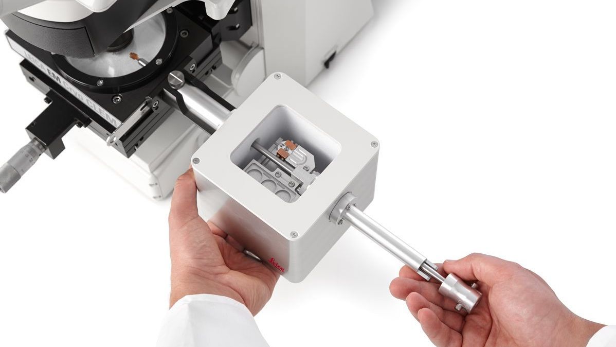
Cosa è importante per la preparazione dei campioni crio-EM?

Il Cryo EM beneficia della vitrificazione mediante congelamento rapido per mantenere le strutture nel loro stato naturale.

Le soluzioni Leica per la crio-preparazione, tra cui la vetrificazione avanzata, il rivestimento, la sezione ultra sottile, la crio-pianificazione e i flussi di lavoro personalizzabili di trasferimento crio e CLEM, consentono di ottenere campioni riproducibili e privi di contaminazione. Contribuiscono a mantenere le strutture native, forniscono un targeting preciso e consentono l'imaging crio EM ad alta risoluzione.

Perché scegliere gli strumenti e le soluzioni Leica per la preparazione dei campioni EM?


Preparazione del campione SEM

Gli strumenti per la preparazione dei SEM di Leica Microsystems includono rivestimenti sputter ad alto vuoto ed essiccatori a punto critico. Queste soluzioni garantiscono immagini di superficie di alta qualità. Inoltre, gli strumenti Leica forniscono risultati coerenti e affidabili per i vostri studi SEM.


Preparazione del campione TEM

I ricercatori possono affidarsi alle soluzioni di preparazione TEM di Leica Microsystems per ottenere precisione e affidabilità. Strumenti essenziali come gli ultramicrotomi e i sistemi di colorazione automatizzati aiutano a produrre sezioni ultrasottili e immagini ad alta risoluzione. Lo Sputter coating migliora la conduttività del campione e la qualità delle immagini.


Preparazione Cryo-EM completa

Con gli strumenti di crio-preparazione di Leica Microsystems, tra cui i congelatori a tuffo e i congelatori ad alta pressione, i ricercatori assicurano un congelamento e una vetrificazione rapidi. Inoltre, il nostro sistema di trasferimento dei campioni per i flussi di lavoro criogenici offre opzioni di trasferimento per tutti i tipi di campioni. Questa opzione li protegge dalla contaminazione, garantendo un'analisi Cryo-EM accurata e dettagliata.

Articoli correlati

Leggete i nostri ultimi articoli sui flussi di lavoro per la preparazione dei campioni EM & Utilizzi

Il portale informativo di Leica Microsystems offre materiale didattico e di ricerca scientifica su vari temi della microscopia. Il contenuto è stato progettato per aiutare i principianti, i professionisti e gli scienziati nel lavoro quotidiano e negli esperimenti.

Altri articoli
Sectioning resin-embedded sambles

Ultramicrotomy eBook: Targeting, Trimming & Alignment

Ultramicrotomy is evolving rapidly, and today’s microscopes demand high‑quality sections, precise targeting, and reproducible workflows. This eBook brings together expert application notes, automated…
Foraminifera (Ammonia confertitesta) 
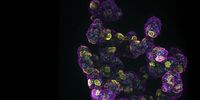
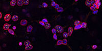
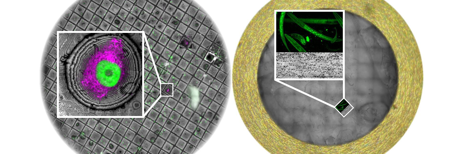
labeled with membrane-permeable calcein, high-pressure frozen in salt water using EM ICE. The sample was cryo-planed and targeted with the M205 on the Cryo-Fluo Enuity, then transferred under cryo conditions to the Cryo-Stellaris for widefield and confocal imaging, revealing details of the staining pattern. Image courtesy: David Evans, University of Southampton.

High-Pressure Freezing for Organoids: Cryo CLEM & FIB Lift Out

Master cryo EM workflow steps for challenging 3D samples: when to choose HPF vs. plunge freezing, reproducible blotting/ice control, contamination aware transfers, Cryo CLEM 3D targeting in organoids,…
Electron microscope (EM) 
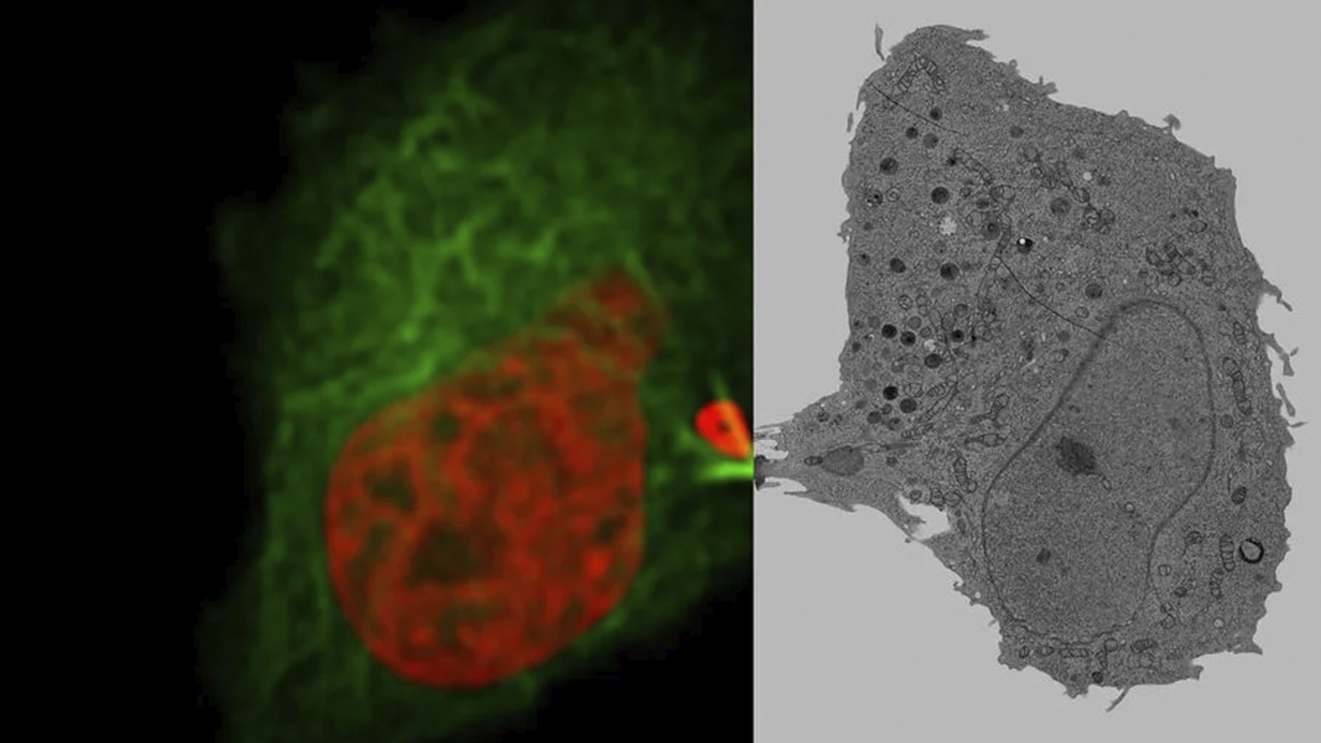
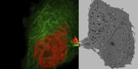
image of a cross section of C. elegans (roundworm). Courtesy of T. Müller-Reichert, MPI-CBG, Dresden, Germany and K. McDonald, University of California, Berkeley, USA.

Brief Introduction to High-Pressure Freezing for Cryo-Fixation

Preparation of biological specimens for electron microscopy (EM) often requires cryo-fixation which does not introduce significant structural alterations of cellular constituents. A common method used…
TEM micrographs of polymer sections. Left: Poly(styrene)-
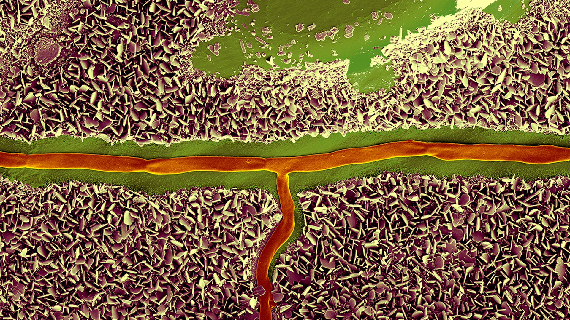
b-poly(isoprene). Right: Poly(styrene)-b-poly(methyl methacrylate).

Ultramicrotome Sectioning of Polymers for TEM Analysis

We demonstrate the capabilities of the UC Enuity ultramicrotome from Leica Microsystems for preparing ultrathin sections of polymer samples under both ambient and cryogenic conditions. By presenting…
Final Segmentation of organelles in Trichomonas species. Magenta – costa, light blue – hydrogenosomes, turquoise – ER, red – vacuoles, yellow – axostyle, green – Golgi apparatus.  Sample courtesy of Isabelle Guerin-Bonne, Low Kay En, Electron Microscopy Unit, Yong Loo Lin School of Medicine, National University of Singapore. Scale bar: 1 µm.

Volume EM and AI Image Analysis

The article outlines a detailed workflow for studying biological tissues in three dimensions using volume-scanning electron microscopy (volume-SEM) combined with AI-assisted image analysis. The focus…

Integrated Serial Sectioning and Cryo-EM Workflows for 3D Biological Imaging

This on-demand webinar explores how integrated tools can support electron microscopy workflows from sample preparation to image analysis. Experts Andreia Pinto, Adrian Boey, and Hoyin Lai present the…
SEM image of the full Li-NMC electrode sample, showing the two porous layers and the metal film at the center of the structure.

Revealing Sodium Battery Degradation via Cryo-EM and CryoFIB

Explore how cryogenic electron microscopy and focused ion beam techniques uncover the intrinsic structure of sodium battery interfaces. This webinar presents a new degradation model based on separator…
Mouse hippocampus brain slice on a grid after HPF using the “Waffle Method”.

The “Waffle Method”: High-Pressure Freeze Complex Samples

This article describes the advantages of a special high pressure freezing method, the so-called “Waffle Method”. Learn how the “Waffle Method” uses EM grids as spacers for high-pressure freezing,…
UC Enuity with Diatome diamond knife.

Mastering Polymer Sectioning with Helmut Gnaegi

When it comes to ultramicrotomy, few names carry the weight of Helmut Gnaegi. As co-founder of Diatome, a global leader in diamond knife technology, Helmut has spent decades refining the art and…
C. elegans embedded in Lowicryl® HM20; pharynx showing red fluorescence (mCherry). The overview shows a front view onto the resin capsule formed by the bottom of a flow-through chamber of the EM AFS2. The capsule was pretrimmed manually. The blockface was trimmed automatically using the AutoTrim function of UC Enuity guided by fluorescence of the worm. Edge length of both squares in relation to the images is 250 µm.

How Fluorescence Guides Sectioning of Resin-embedded EM Samples

Electron microscopes, including transmission electron microscopes (TEM) and scanning electron microscopes (SEM), are widely utilized to gain detailed structural information about biological samples or…
14: 7-day old 3D cell culture of MDCK cells stably transfected with; Mx1-GFP (green), SPY555-Actin (yellow) and WGA-AlexaTM 647 (magenta), and imaged using a THUNDER Imager Cell Spinning Disk. Shown are both the spinning disk image (left) and the same image after THUNDER Computational Clearing was applied (right); scale bar 50 μm.

Organoidi e colture cellulari in 3D

Uno dei più entusiasmanti progressi recenti nella ricerca nel campo delle scienze della vita è lo sviluppo di sistemi di coltura cellulare in 3D, quali gli organoidi, gli sferoidi o i modelli di organi-su-un-chip. Una coltura cellulare in 3D è un ambiente artificiale in cui le cellule sono in grado di crescere e interagire con l'ambiente circostante in tutte le 3 dimensioni. Queste condizioni sono simili a come sarebbero in vivo.

PER SAPERNE DI PIÙ

Image of C2C12 cells: The cells are stained with lamin B (magenta) which indicates nuclear structure, Hoechst (blue) indicating DNA, and γH2AX (yellow) indicating damage to DNA. Cells were imaged using a THUNDER Imager 3D Live Cell with a 63X/1.4 oil immersion objective.

Ricerca sulla biologia cellulare

L'imaging di biologia cellulare viene realizzato utilizzando una gamma di microscopi ottici ed elettronici. Le soluzioni di imaging offerte da Leica Microsystems sono progettate per ottimizzare la ricerca sulla biologia cellulare.

PER SAPERNE DI PIÙ

Mammalian cells expressing H2B-mCherry and alphaTubulin-mEGFP to visualize DNA and microtubules respectively. Left: Widefield imaging using THUNDER Imager. Right: Sample after correlated EM imaging using Leica Microsystems Coral Life workflow. Cells were cryo-immobilized by high-pressure freezing 60 min after the onset of cytokinesis. The intercellular bridge was imaged under TEM after freeze substitution and resin infiltration.

Microscopia correlativa per sistemi ottici ed elettronici (CLEM)

Le soluzioni CLEM di Leica Microsystems garantiscono la vitalità dei campioni, la qualità dei campioni e un meccanismo di targeting 3D preciso e affidabile. Gli utenti possono sfruttare queste soluzioni per identificare direttamente la cellula giusta al momento giusto, ottenere dati crio-confocali ad alta risoluzione e inserire le informazioni sulla fluorescenza nel contesto ultrastrutturale.

PER SAPERNE DI PIÙ

Coltura cellulare

Crescere le cellule in laboratorio rappresenta la base per le attività scientifiche nei campi della biologia cellulare, ricerca sul cancro, biologia dello sviluppo e in diversi altri campi di ricerca farmaceutica e di scienze della vita. Venite a scoprire come Leica supporta la coltura di cellule animali in laboratorio.

PER SAPERNE DI PIÙ

14: 7-day old 3D cell culture of MDCK cells stably transfected with; Mx1-GFP (green), SPY555-Actin (yellow) and WGA-AlexaTM 647 (magenta), and imaged using a THUNDER Imager Cell Spinning Disk. Shown are both the spinning disk image (left) and the same image after THUNDER Computational Clearing was applied (right); scale bar 50 μm.
Image of C2C12 cells: The cells are stained with lamin B (magenta) which indicates nuclear structure, Hoechst (blue) indicating DNA, and γH2AX (yellow) indicating damage to DNA. Cells were imaged using a THUNDER Imager 3D Live Cell with a 63X/1.4 oil immersion objective.
Mammalian cells expressing H2B-mCherry and alphaTubulin-mEGFP to visualize DNA and microtubules respectively. Left: Widefield imaging using THUNDER Imager. Right: Sample after correlated EM imaging using Leica Microsystems Coral Life workflow. Cells were cryo-immobilized by high-pressure freezing 60 min after the onset of cytokinesis. The intercellular bridge was imaged under TEM after freeze substitution and resin infiltration.
Scroll to top