Accessori del microscopio

Accessori del microscopio

Leica Microsystems offre una vasta gamma di accessori per microscopi, con soluzioni che si adattano alla perfezione a specifici sistemi di Imaging in termini di esigenze e budget. Consulta il nostro catalogo relativo a illuminazione, obiettivi, cubi portafiltri, ruote portafiltri e molto altro ancora.

Leggi gli articoli più recenti

Il portale informativo di Leica Microsystems offre materiale didattico e di ricerca scientifica su vari temi della microscopia. Il contenuto è stato progettato per aiutare i principianti, i professionisti esperti e gli scienziati nel lavoro quotidiano e negli esperimenti.

A Guide to Polarized Light Microscopy

Polarized light microscopy (POL) enhances contrast in birefringent materials and is used in geology, biology, and materials science to study minerals, crystals, fibers, and plant cell walls.

Factors to Consider when Selecting Clinical Microscopes

What matters if you would like to purchase a clinical microscope? Learn how to arrive at the best buying decision from our Science Lab Article.

Clinical Microscopy: Considerations on Camera Selection

The need for images in pathology laboratories has significantly increased over the past few years, be it in histopathology, cytology, hematology, clinical microbiology, or other applications. They serve many purposes on top of the documentation of the diagnosis. Yet, the view through the eyepieces and the image are different in nature, the one is an optical image, the other a digital image. Looking at a few aspects of this process that are related cameras will help you make sure you can obtain the images with all the detail and color fidelity you need.

How to Save Time and Samples by Automated Ultramicrotomy

This article describes how 3D micro-CT data of a resin-embedded electron microscopy sample can be used to trim the specimen down to a defined target plane prior to sectioning. The interactive and automated approach described using Leica Microsystems’ UC Enuity saves time, reduces sample loss and training time for unexperienced users.

Designing the Future with Novel and Scalable Stem Cell Culture

Visionary biotech start-up Uncommon Bio is tackling one of the world’s biggest health challenges: food sustainability. In this webinar, Stem Cell Scientist Samuel East shows how they make stem cell culture media for cellular agriculture safe and economically viable. See how they achieved a 1000x reduction in media costs and developed animal free, food safe iPSC culture media.

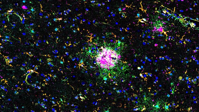
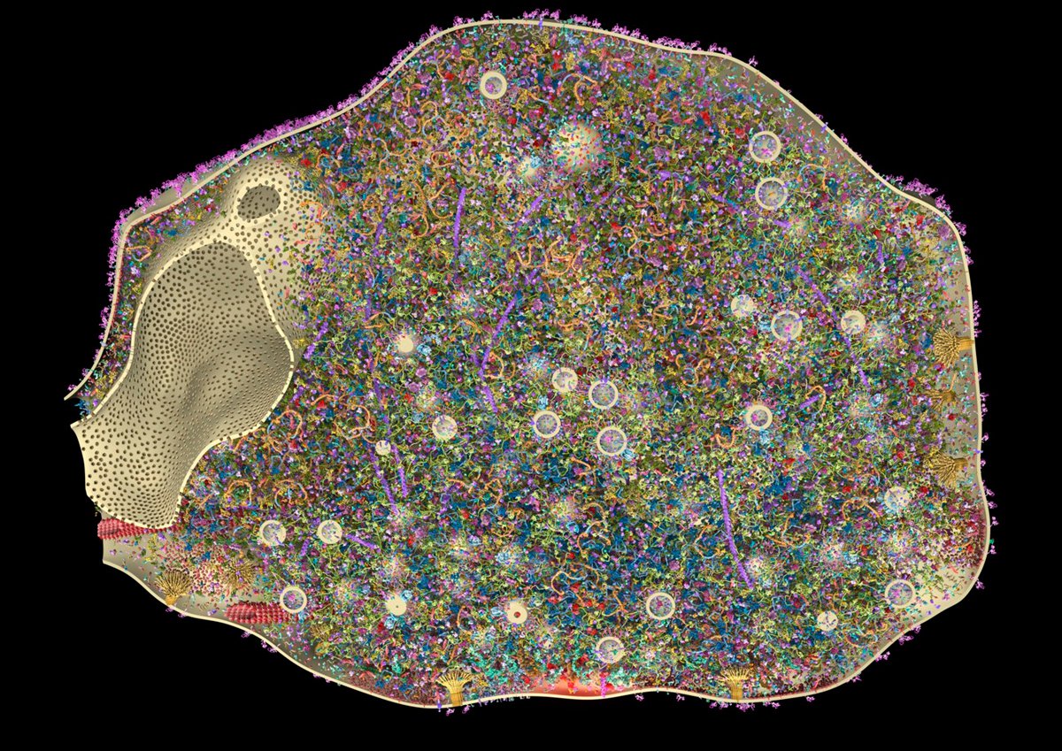
Explore Alzheimer's Spatial Proteome with Big Data

Alzheimer's disease, a genetic and sporadic neurodegenerative condition, leads to cognitive decline in mid to late life, marked by β-amyloid plaques and tau tangles. With limited treatment options, new investigative strategies are crucial. The Cell DIVE multiplexed imaging solution allows examination of Alzheimer's brain tissue, potentially uncovering new research avenues. Here, we showcase the Cell DIVE image viewer, enabling users to access the full Alzheimer's multiplexed dataset directly in their browser.

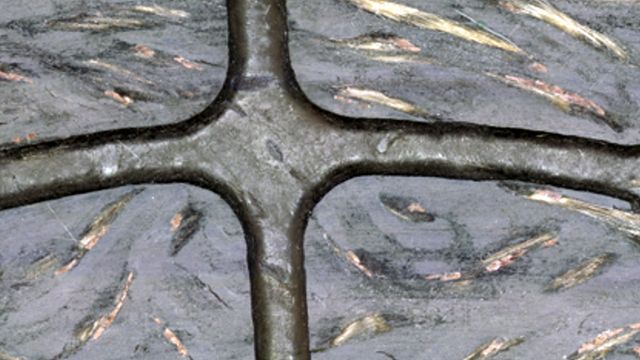
Automotive Part Verification and Development according to Specifications

Automotive part verification during the development and production of parts and components by suppliers or manufacturers is important for ensuring that specifications are met. Specifications are critical for maintaining performance standards and safe operation of vehicles. However, there is always demand for efficient and cost-effective R&D and production while meeting ever-stricter quality standards. This article shows how investigation and documentation of auto parts and components to verify specifications can be done easily and quickly with digital microscopy.

Mica: A Game-changer for Collaborative Research at Imperial College London

This interview highlights the transformative impact of Mica at Imperial College London. Scientists explain how Mica has been a game-changer, expanding research possibilities and facilitating interdisciplinary collaboration. They explain how detailed live cell imaging with Mica provides more meaningful information, keeping scientists at the forefront of research. The team foresees Mica continuing to open new research avenues, including the study of microfluidics and other advanced applications.

From Bench to Beam: A Complete Correlative Cryo Light Microscopy Workflow
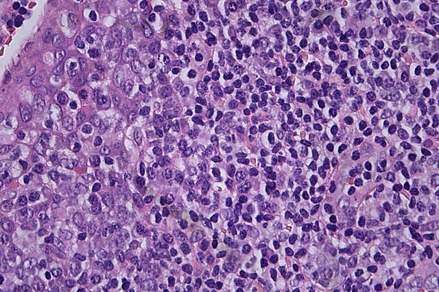
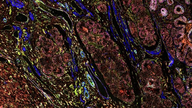
In the webinar entitled "A Multimodal Vitreous Crusade, a Cryo Correlative Workflow from Bench to Beam" a team of experts discusses the exciting world of correlative workflows for structural biology that empower researchers to study fine details of biological structures. Watch and explore the latest developments, instruments, and techniques in cryo workflows for correlative light electron microscopy (cryo-CLEM).
Feb 06, 2025
Webinar
CLEM

Ricerca sul cancro

Il cancro è una malattia complessa ed eterogenea causata da cellule che hanno dei difetti nella regolazione della crescita. I cambiamenti genetici ed epigenetici in una cellula o in un gruppo di cellule alterano il normale funzionamento e provocano una crescita e una proliferazione cellulare autonoma e incontrollata.

Depth of Field in Microscope Images

For microscopy imaging, depth of field is an important parameter when needing sharp images of sample areas with structures having significant changes in depth. In practice, depth of field is determined by the correlation between numerical aperture, resolution, and magnification. For the best possible visualization of samples, modern microscopes can be adjusted to produce an optimum balance between depth of field and resolution. In theory, these parameters are inversely correlated.

Dive into Pancreatic Cancer Research with Big Data

Pancreatic cancer, with a mortality rate near 40%, is challenging to treat due to its proximity to major organs. This story explores the complex biology of pancreatic ductal adenocarcinoma (PDAC), examining molecular and spatial determinants of tumor aggression in metabolism, apoptosis, and immunity. Access the full Cell DIVE dataset in your browser to delve deeper into these findings.

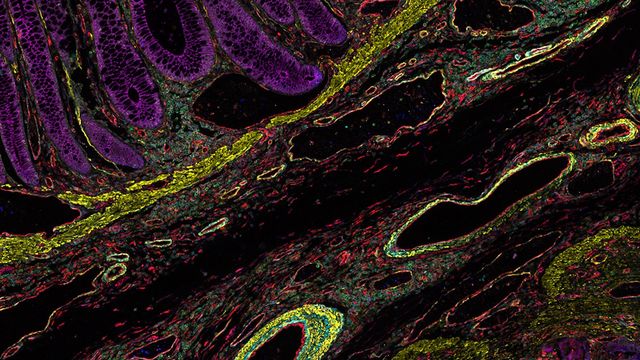
Uncover the Hidden Complexity of Colon Cancer with Big Data

Colorectal cancer poses a significant health burden. While surgery is effective initially, some patients develop recurrent secondary disease with poor prognosis, necessitating advanced therapies like immunotherapies. Spatial biology approaches, such as multiplexed imaging with Cell DIVE, can provide crucial insights for developing novel treatments. Access the full Cell DIVE dataset in your browser to explore these findings further through the Minerva image viewer.

Introduction to 21 CFR Part 11 for Electronic Records of Cell Culture

This article provides an introduction to the recommendations of 21 CFR Part 11 from the FDA, specifically focusing on the audit trail and user management in the context of cell-culture laboratories. It is intended for professionals in the biotechnology and pharmaceutical industries who are responsible for ensuring agreement with 21 CFR part 11 for electronic records and electronic signatures. A digital microscope approach, e.g. with Mateo FL, offers the advantage of more consistent and efficient electronic documentation of cell-culture results compared to a paper-based method.

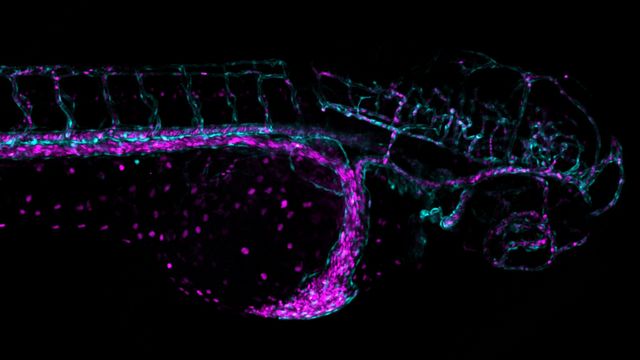
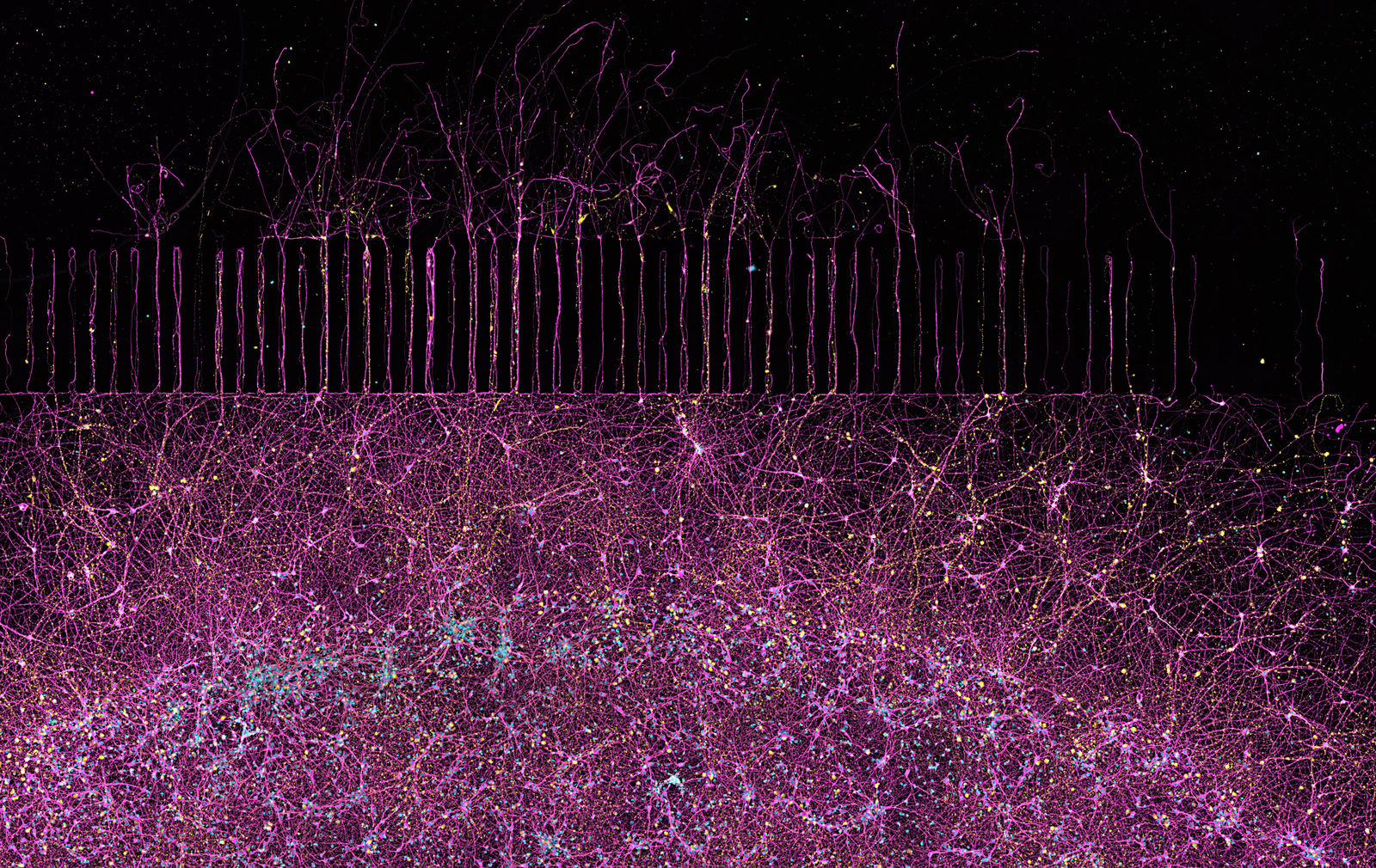
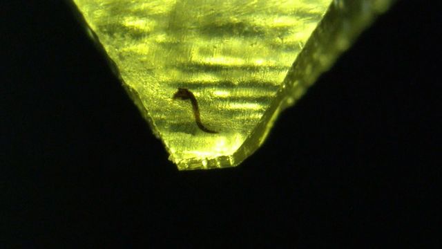
Overcoming Challenges with Microscopy when Imaging Moving Zebrafish Larvae

Zebrafish is a valuable model organism with many beneficial traits. However, imaging a full organism poses challenges as it is not stationary. Here, this case study shows how zebrafish larvae can be imaged during stationary periods and easily relocated after movement. The seamlessly integrated widefield and confocal capability of Mica is leveraged to capture fast events, like the heartbeat, with virtually no out-of-focus background noise which is inherent to standard widefield systems.

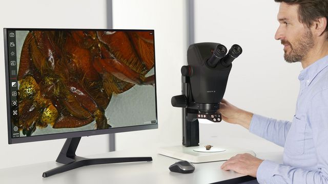
Microscopi da dissezione
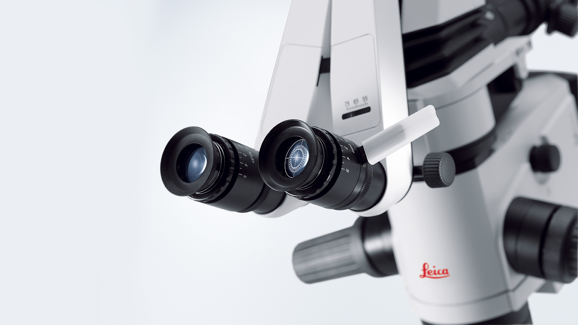
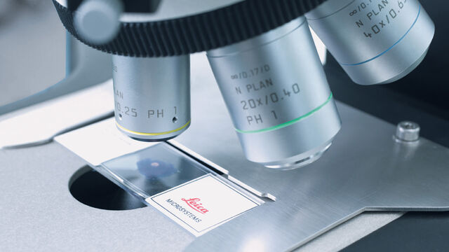
Ogni volta che devono essere eseguite delle dissezioni, possono essere necessarie molte ore di osservazione attraverso gli oculari di un microscopio da dissezione. Leica Microsystems ti offre una vasta scelta di microscopi e una gamma completa di componenti e accessori per microscopi da dissezione, per soddisfare ogni tua esigenza.

Oculari e reticoli

Per i tubi del microscopio è disponibile un'ampia scelta di oculari con ingrandimento 10x, 12,5x, 16x o 25x (per valori di campo fino a 25 mm). Sono disponibili oculari speciali per chi indossa occhiali da vista e oculari con lenti regolabili (lenti M) progettate per adattarsi a diversi reticoli.

Gli oculari standard

Gli oculari standard hanno ingrandimento 10x; gli oculari con ingrandimento 16x e 25x sono destinati esclusivamente ad applicazioni speciali. Tutti gli oculari sono dotati di paraocchi rimovibili o ripiegabili, pertanto possono essere utilizzati con o senza occhiali.

Gli oculari identificati con la lettera M sono dotati di lenti di messa a fuoco per l'equalizzazione diottrica (da -6,8 a +4,2 o da -6 a +5) e portareticolo. Il diametro esterno degli oculari è D = 30 mm.

Il diametro del reticolo è D = 26 mm. Le caratteristiche tecniche sono incise sugli oculari, ad esempio HC PLAN 10x/20 M. HC PLAN = tipo di correzione, 10x = ingrandimento/20 = numero di campo FOV, = per chi indossa occhiali (pupilla di uscita di grandi dimensioni), M = regolazione diottrica/portareticolo.

Oculari con FOV 20

Show details
  • Oculare HC PLAN 10x/20 BR
    11 507 801
  • Oculare HC PLAN 10x/20 BR.M
    11 507 802

Oculare con FOV 22

Show details
  • Oculare HC PLAN S 10x/22 Br.M
  • 11507820

Oculare con FOV 25

Show details
  • Oculare HC PLAN S 10x/25 Br.M
    11 507 808

Oculari speciali a elevato ingrandimento

Show details
  • Oculare HC PLAN 12.5x/16 BR.M
    11 506 515
  • Oculare 16x/14B, regolabile
    10 445 301
  • Oculare 25x/9.5B, regolabile
    10 445 302
  • Anello distanziatore per oculari 16x/14B e oculare 25x/9.5B
    11 506 808

Reticoli di messa a fuoco e di delimitazione

Show details

Per misurazioni di lunghezza, metodi di confronto e di calcolo (Ø = 26 mm)

  • Reticolo 10 mm = 100 parti
    11 506 950
  • Reticolo 10 mm = 200 parti
    11 506 951
  • Reticolo a croce
    11 506 953
  • Reticolo a croce con graduazione, 10 mm = 100 parte
    11 506 952
  • Reticolo con griglia 10 x 10 mm, 0,1 mm graduazione
    11 506 954
  • Reticolo con griglia 10 x 10 mm, 1,0 mm graduazione
    11 506 955

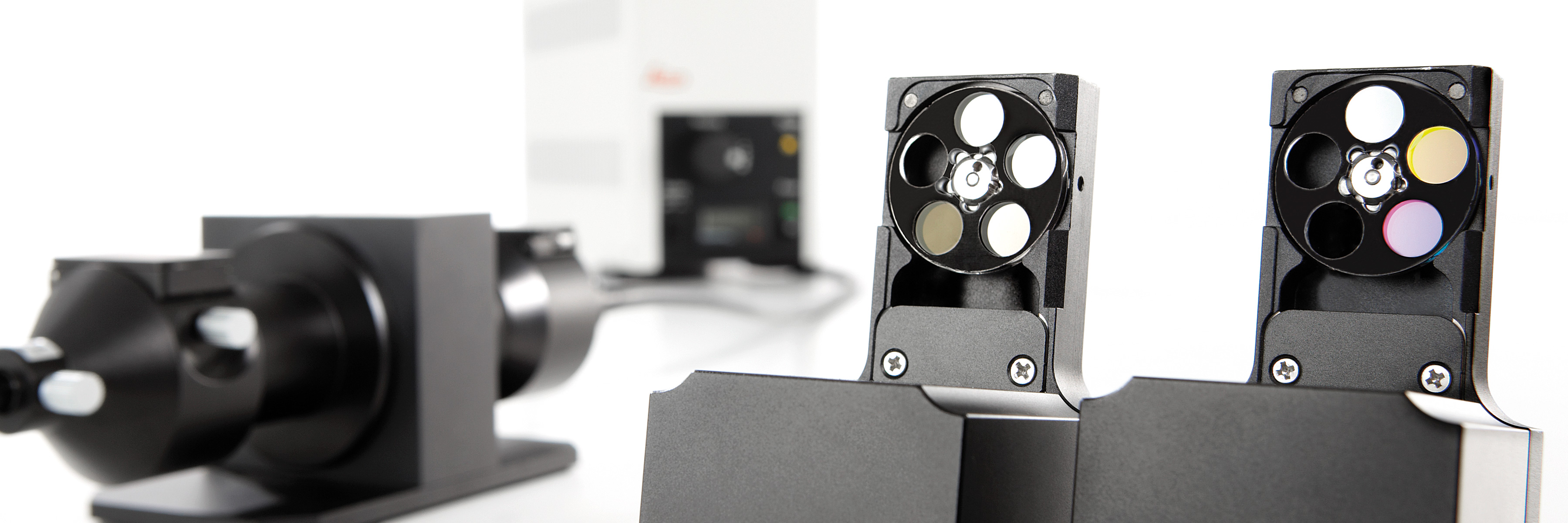
Fluorescenza ad alta velocità: ruote portafiltri esterne per applicazioni su cellule vive

Sono rapide e prive di vibrazioni grazie alle loro dimensioni ridotte, e offrono una versatilità eccezionale. Queste straordinarie caratteristiche sono tipiche delle ruote portafiltri veloci di Leica Microsystems per il controllo di eccitazione, attenuazione ed emissione. La rapidità di commutazione le rende adatte anche alle applicazioni più complesse, come l'Imaging FRET o CA++ (Fura2).

  • Controllo di eccitazione, attenuazione ed emissione ad alta velocità
  • Tempo di commutazione 24 ms (posizione adiacente)
  • Vibrazioni minime
  • Struttura piccola e compatta
  • Controllo ad alta velocità sincronizzato tramite potenti soluzioni hardware e software (Leica AF6000 E, AF6000, AF6500 e AF7000)
  • Velocità di acquisizione: 31 fps
  • Flessibile con diverse configurazioni: dischi a cinque posizioni con filtri
  • Cursore motorizzato inseribile nell'adattatore del Leica EL6000, nello stativo del microscopio o nello speciale attacco C per il controllo dell'emissione
  • Possono essere adattati contemporaneamente fino a quattro cursori
  • Pacchetti applicativi predefiniti per fluorescenza standard, Fura2 e FRET

Brochure External Filter Wheels, EN

Le ruote portafiltri ultra veloci di Leica Microsystems garantiscono un Imaging accurato

I filtri per fluorescenza sono leggeri, hanno diametro ridotto, consentono una commutazione estremamente rapida tra filtri e producono vibrazioni minime.

Eccitazione ed emissione di luce possono essere selezionati in appena 24 millisecondi (velocità di acquisizione: 31 fps). Altrettanto rapidamente possono essere attenuati i singoli colori d'eccitazione.

Sono disponibili pacchetti applicativi personalizzati, anche per esperimenti estremamente complessi nel campo delle Life Sciences. Il controllo software si realizza attraverso Leica Application Suite o la Serie Leica AF, che offrono entrambe un'interfaccia utente ottimizzata che guida gli utenti anche negli esperimenti più complicati.

Inoltre, Leica offre una dotazione completa di fotocamere digitali. Insieme alla sorgente di luce esterna priva di allineamento Leica EL6000, è possibile ottimizzare ulteriormente il tempo di risoluzione dei segnali di fluorescenza.

Settore Medico

Nelle applicazioni mediche, Leica vi supporta con sistemi ottici di alta qualità che vi offre una visualizzazione nitida, luminosa ed uniforme.

Per saperne di più

Microscopi per la ricerca Life Sciences

I microscopi per la ricerca Life Sciences di Leica Microsystems supportano le esigenze di imaging della comunità scientifica con la massima capacità innovativa e la competenza tecnica.

Per saperne di più

Applicazioni industriali

Grazie a una vasta gamma di soluzioni e un servizio assistenza fornito da esperti locali, con i microscopi Leica Microsystems per l'industria e la produzione è possibile ridurre al minimo l'inattività.

Per saperne di più

Settore Medico

Nelle applicazioni mediche, Leica vi supporta con sistemi ottici di alta qualità che vi offre una visualizzazione nitida, luminosa ed uniforme.

Per saperne di più

Microscopi per la ricerca Life Sciences

I microscopi per la ricerca Life Sciences di Leica Microsystems supportano le esigenze di imaging della comunità scientifica con la massima capacità innovativa e la competenza tecnica.

Per saperne di più

Scroll to top