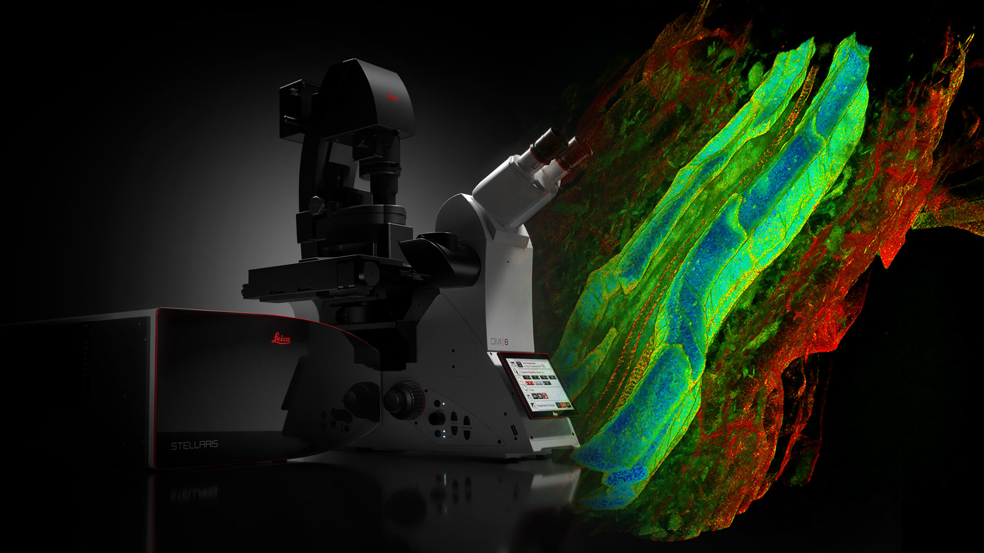
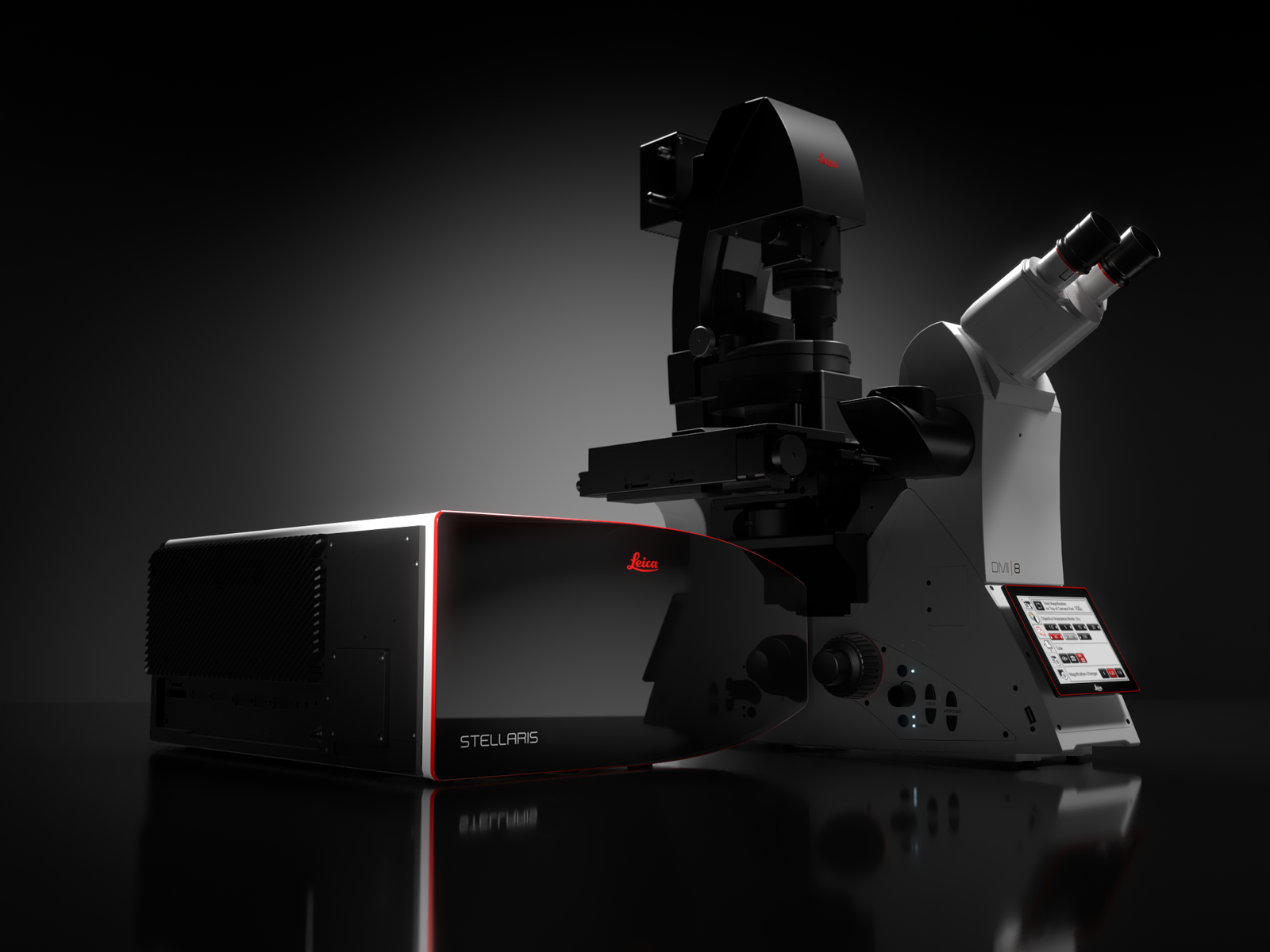
STELLARIS Piattaforma di microscopia confocale
STELLARIS di nuova generazione è una piattaforma per microscopi confocali progettata in base alle tue esigenze di ricerca.
I microscopi confocali STELLARIS possono essere combinati con tutte le modalità Leica, tra cui FLIM, STED, MP, DLS e CRS. La piattaforma confocale STELLARIS di nuova generazione è la tua corsia preferenziale per una ricerca più efficace. Power. Potential. Productivity.
The Potential to discover more
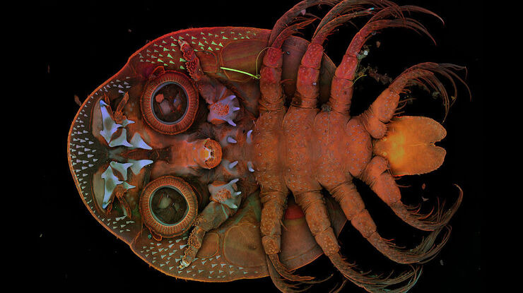
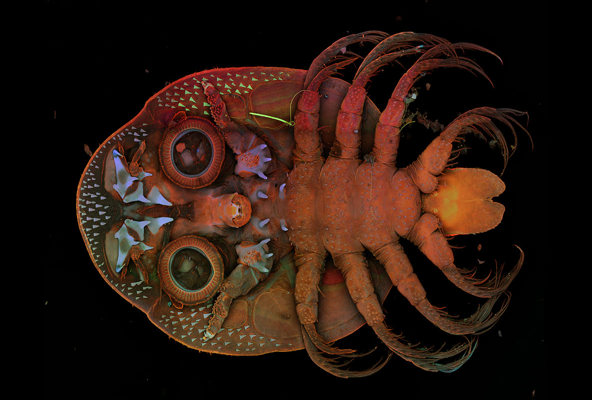
L'esclusiva tecnologia TauSense di STELLARIS consente di estrarre un ulteriore livello di informazioni da ogni campione e aumentare l'impatto scientifico della ricerca. TauSense comprende strumenti di imaging applicativi basati sul tempo di vita della fluorescenza che possono essere utilizzati per esplorare la funzione delle molecole nel contesto cellulare.
- TauContrast assicura un accesso immediato a informazioni funzionali quali lo stato metabolico, il pH e la concentrazione ionica.
- TauGating migliora la qualità delle immagini rimuovendo i contributi di fluorescenza indesiderati.
- TauSeparation aiuta a espandere la combinazione di segnali fluorescenti nell'esperimento in aggiunta alle opzioni di separazione spettrali.
- TauInteraction consente di rilevare e quantificare in modo semplice le interazioni molecolari (ad esempio l'interazione proteina-proteina).
Accesso ai dati che contano
Ottieni risultati di alta qualità più rapidamente con la Autonomous Microscopy di Aivia.
- Il workflow di rilevamento di eventi rari basato sull'IA per life science su STELLARIS rileva autonomamente fino al 90% degli eventi rari nei campioni biologici.
- Riduci radicalmente il tempo di acquisizione dati fino al 70%, poiché gli oggetti di interesse vengono identificati e registrati in modo esclusivo. Registra solo ciò di cui hai bisogno, risparmiando spazio prezioso sul tuo disco rigido.
- Trascorri meno tempo al microscopio: il workflow autonomo per il rilevamento di eventi rari richiede solo una frazione del tempo solitamente necessario.
- Grazie alla Autonomous Microscopy di Aivia potrai condurre esperimenti che in precedenza non erano possibili a causa di vincoli di tempo e complessità.
STELLARIS con WLL
STELLARIS è l'unico sistema confocale con WLL integrato, combinato con il nostro beam splitter acusto-ottico (AOBS) brevettato e la gamma di rilevatori Power Hyd. Unitamente all'esclusiva tecnologia TauSense, STELLARIS definisce un nuovo standard per la qualità delle immagini e per la quantità di informazioni generate.
STELLARIS con laser a luce bianca può essere combinato con FAst Lifetime CONtrast (FALCON), STED, Deep In Vivo Explorer (DIVE), Digital Light Sheet (DLS), Cryo e CARS. STELLARIS massimizza il potenziale di queste modalità e dà la possibilità e il potenziale di fissare nuovi standard per la ricerca.
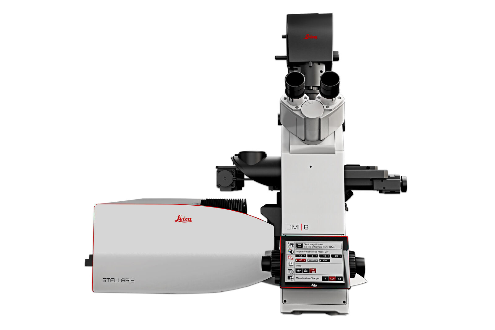
STELLARIS
STELLARIS offre rivelazione spettrale, rivelatori a conta fotonica e una super risoluzione confocale.
L'imaging confocale multicanale è facilmente realizzabile grazie all'interfaccia utente smart, ImageCompass, che ti guida in modo intuitivo attraverso la configurazione e l'acquisizione dell'esperimento.
The Productivity to do more
Configurazione e acquisizione semplificate: ImageCompass, l'interfaccia utente smart di STELLARIS, offre agli utenti un modo semplice e intuitivo per configurare anche gli esperimenti più complessi in pochi clic.
- Semplicemente veloce: eccellente qualità dell'immagine in tempo reale e alla massima velocità con la super-risoluzione LIGHTNING, Dynamic Signal Enhancement (DSE) di Aivia.
- Interfaccia utente intuitiva: ImageCompass ti guida dalla configurazione dell'esperimento fino all'acquisizione.
- Ottimizza il tuo esperimento: Strumenti come LAS X Navigator sono perfettamente integrati per un imaging semplice.
The power to see more
La famiglia di detector Power HyD offre una maggiore photon detection efficiency (PDE)*, un dark noise estremamente basso e un intervallo di rilevamento spettrale da 410 a 850 nm.
- Migliorata qualità delle immagini: combinazione ideale di luminosità, risoluzione e contrasto.
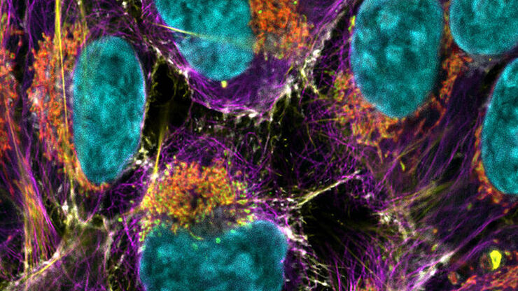
- Libertà spettrale completa: i nostri laser a luce bianca di ultima generazione consentono di utilizzare contemporaneamente fino a 8 linee di eccitazione singole in tutto lo spettro. È possibile acquisire un maggior numero di combinazioni di fluorofori rispetto a qualsiasi altra piattaforma confocale e utilizzare più fluorocromi in parallelo, ampliando le opzioni nella regione NIR.
- Live cell imaging delicato: Osserva il campione e preservane l'integrità per periodi di tempo più lunghi grazie all'acquisizione efficiente del segnale con livelli di illuminazione più bassi
*Photo Detection Efficiency (PDE) 2 volte superiore rispetto ai tradizionali fotomoltiplicatori (PMT) multi-alcalini e 3 volte superiore nella zona estesa del rosso.