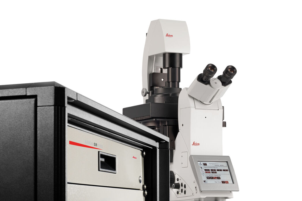
SR GSD 3D
倒立顕微鏡
光学顕微鏡
製品紹介
Home
Leica Microsystems
SR GSD 3D 3D 局在化顕微鏡超解像システム
The Evolution of Resolution
アーカイブした製品
This item has been phased out and is no longer available. Please contact us to enquire about recent alternative products that may suit your needs.
細胞プロセスの正確な局在化を可視化することは、分子構造と機能の相互作用を理解する上できわめて重要です。 ライカ SR GSD 3D 超解像顕微鏡は、GSD(Ground State Depletion)または dSTORM(Direct STochastical Optical Reconstruction Microscopy)技術に基づき、2D だけでなく 3D の超解像イメージングも可能にし、最高の精度、再現性、そして現在のWidefield顕微鏡では最大分解能を実現しています。 ライカ SR GSD 3D は自動化された TIRF システムをベースとしています。 この多機能なシステムは、研究者が自由に、生細胞における各自のアプリケーションや先進の蛍光イメージングに適合する自由度が確保されています。
Alexa 647-Ab を用いてVimentinを染色したヒト内皮細胞(Huvec)3D 画像。Medium: gloxy-like buffer 資料提供: K. Jalink and L. Nahidi Azar, Amsterdam, The Netherlands.

