Cell DIVE
광학 현미경
제품소개
홈
Leica Microsystems
Cell DIVE 멀티플렉스 이미징 솔루션
개방형 멀티플렉스로 조직 연구 혁신하기.
최신 기사를 읽어 보세요

History, Developments and Trends of Microscopy in Cancer Research
Cancer is a global disease, with 18 million new cases diagnosed and 10 million cancer-related deaths worldwide in 2020. This burden is set to increase, with a projected increase in cases of ~55% by…
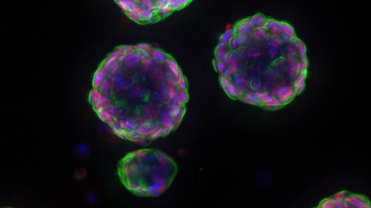
 and astrocytes (green) in a cortical spheroid derived from human induced pluripotent stem cells.")
Guide to Live-Cell Imaging
For a wide range of applications in various research fields of life science, live-cell imaging is an indispensable tool for visualizing cells in a state as close to in vivo, i.e. living and active, as…

Factors to Consider When Selecting a Research Microscope
An optical microscope is often one of the central devices in a life-science research lab. It can be used for various applications which shed light on many scientific questions. Thereby the…
.")
AI-Powered Hi-Plex Spatial Analysis Tools for Breast Cancer Research
Breast cancer (BC) is the leading cause of cancer-related deaths in women. Investigating the tumor microenvironment (TME) is crucial to elucidate the mechanisms of tumor progression. Systematic…

Multiplexed Imaging Reveals Tumor Immune Landscape in Colon Cancer
Cancer immunotherapy benefits few due to resistance and relapse, and combinatorial therapeutic strategies that target multiple steps of the cancer-immunity cycle may improve outcomes. This study shows…

신경과학 연구
신경변경 질환에 대해 더 잘 이해하기 위해 노력하고 있거나 신경계 기능을 연구하고 계십니까? 라이카마이크로시스템즈의 이미지 솔루션을 통해 발전을 이룰 수 있는 방법을 알아보세요.

Boston and San Francisco Innovation Hubs
Boston and San Francisco Innovation Hubs are here to help you advance scientific discovery. We provide researchers access to state-of-the-art microscope technology and expert guidance. Located in the…

Transforming Research with Spatial Proteomics Workflows
Spatial Proteomics, Nature Methods 2024 Method of the Year, is driving research advancements in cancer, immunology, and beyond. By combining positional data with high throughput imaging of proteins in…
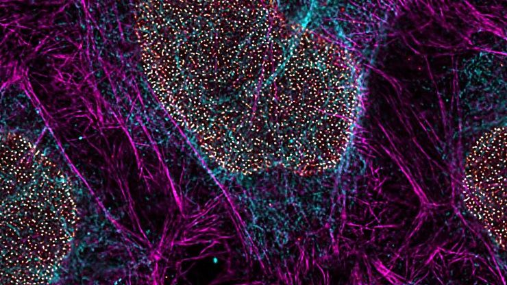
, microglia (TMEM119, IBA1), and Alzheimer’s-associated markers (β-amyloid and p-Tau217).")
Explore Alzheimer's Spatial Proteome with Big Data
Alzheimer's disease, a genetic and sporadic neurodegenerative condition, leads to cognitive decline in mid to late life, marked by β-amyloid plaques and tau tangles. With limited treatment options,…

Uncover the Hidden Complexity of Colon Cancer with Big Data
Colorectal cancer poses a significant health burden. While surgery is effective initially, some patients develop recurrent secondary disease with poor prognosis, necessitating advanced therapies like…

Dive into Pancreatic Cancer Research with Big Data
Pancreatic cancer, with a mortality rate near 40%, is challenging to treat due to its proximity to major organs. This story explores the complex biology of pancreatic ductal adenocarcinoma (PDAC),…

Mapping Tumor Immune Landscape with AI-Powered Spatial Proteomics
Spatial mapping of untreated tumors provides an overview of the tumor immune architecture, useful for understanding therapeutic responses. Immunocompetent murine models are essential for identifying…

Spatial Analysis of Neuroimmune Interactions in Alzheimer’s Disease
Alzheimer’s disease (AD) is a complex neurodegenerative disorder characterized by neurofibrillary tangles, β-amyloid plaques, and neuroinflammation. These dysfunctions trigger or are exacerbated by…

A Guide to Spatial Biology
What is spatial biology, and how can researchers leverage its tools to meet the growing demands of biological questions in the post-omics era? This article provides a brief overview of spatial biology…

Probing Human Alzheimer's Cortical Section using Spatial Multiplexing
Alzheimer’s disease (AD) is the most common neurodegenerative disease and is characterized by the progressive decline of cognitive function. Spatial profiling of AD brain may reveal cellular…

Empowering Spatial Biology with Open Multiplexing and Cell DIVE
Spatial biology and multiplexed imaging workflows have become important in immuno-oncology research. Many researchers struggle with study efficiency, even with effective tools and protocols. Here, we…
 tissue.")
AI-Powered Multiplexed Image Analysis to Explore Colon Adenocarcinoma
In this application note, we demonstrate a spatial biology workflow via an AI-powered multiplexed image analysis-based exploration of the tumor immune microenvironment in colon adenocarcinoma.

A Meta-cancer Analysis of the Tumor Spatial Microenvironment
Learn how clustering analysis of Cell DIVE datasets in Aivia can be used to understand tissue-specific and pan-cancer mechanisms of cancer progression
 tissue.")
Mapping the Landscape of Colorectal Adenocarcinoma with Imaging and AI
Discover deep insights in colon adenocarcinoma and other immuno-oncology realms through the potent combination of multiplexed imaging of Cell DIVE and Aivia AI-based image analysis

IBEX, Cell DIVE, and RNA-Seq: A Multi-omics Approach to Follicular Lymphoma
In a recent study by Radtke et al., a multi-omics spatial biology approach helps shed light on early relapsing lymphoma patients

Accelerating Discovery for Multiplexed Imaging of Diverse Tissues
Explore IBEX: Open-source multiplexed imaging. Join the collaborative IBEX Imaging Community for optimized tissue processing, antibody selection, and human atlas construction.

Transforming Multiplexed 2D Data into Spatial Insights Guided by AI
Aivia 13 handles large 2D images and enables researchers to obtain deep insights into microenvironment surrounding their phenotypes with millions of detected objects and automatic clustering up to 30…

Understanding Tumor Heterogeneity with Protein Marker Imaging
Explore tumor heterogeneity and immune cell dynamics. See how quantitative imaging analysis reveals spatial relationships and molecular insights crucial for advancing cancer research and therapeutics.

The Shape of the Brain: Spatial Biology of Alzheimer’s Disease
Uncover cell identity and brain structure in Alzheimer's disease with Cell DIVE multiplexed imaging, demonstrating how spatial biology can lead to advances in therapy development for…

Discover how Multiplexed Bioimaging can Advance Cancer Research
Explore multiplexing with up to 60 biomarkers, enabling advanced tumor imaging approaches to gather precise, spatially-resolved single-cell data that helps enhance cancer research and clinical…

Spatial Biology: Learning the Landscape
Spatial Biology: Understanding the organization and interaction of molecules, cells, and tissues in their native spatial context

Dig Deeper Into the Complexities of Pancreatic Cancer with Multiplex Imaging
Cell DIVE is an iterative staining workflow for multiplexed imaging that unveils biological pathways to dig deeper into the complexities of pancreatic cancer.

Complex Made Simple: Antibodies in Multiplexed Imaging
Build panels, plan studies, and get the most from precious reagents using this antibody multiplexing guide from Leica Microsystems

Multiplexed Imaging Types, Benefits and Applications
Multiplexed imaging is an emerging and exciting way to extract information from human tissue samples by visualizing many more biomarkers than traditional microscopy. By observing many biomarkers…

Be Confident in your Results with Cell DIVE Validated Antibodies
The Cell DIVE System includes a carefully curated list of hundreds of commercially available antibodies validated to offer optimal specificity and sensitivity in multiplexed imaging. That validation…

암 연구
암은 성장 통제에 결함이 있는 세포에 의해 발생하는 복잡하고 이질적인 질병입니다. 하나 또는 한 그룹의 세포에서 일어나는 유전적 또는 후생적 변화가 정상적인 기능을 방해하고, 자율적이고 통제되지 않는 세포 성장과 증식을 초래합니다
Biopharma
For biopharma, Leica solutions help accelerate drug discovery, enhance cellular analysis, and support data integrity that meets regulations.
세포 분석
세포 분석을 위한 고급 현미경 이미징 솔루션과 AI 기반 분석 소프트웨어는 연구자들이 세포 기능과 세포 하부 구조에 대한 심층적인 인사이트를 얻을 수 있도록 도와줍니다.

고급 조직 이미징 및 분석
Leica Microsystems의 첨단 이미징 솔루션으로 조직 구조와 기능에 대한 통찰력을 확보하여 공간 생물학 및 질병 메커니즘에 대한 이해를 높이세요.
Fields of Application
세포 분석
세포 분석을 위한 고급 현미경 이미징 솔루션과 AI 기반 분석 소프트웨어는 연구자들이 세포 기능과 세포 하부 구조에 대한 심층적인 인사이트를 얻을 수 있도록 도와줍니다.
고급 조직 이미징 및 분석
Leica Microsystems의 첨단 이미징 솔루션으로 조직 구조와 기능에 대한 통찰력을 확보하여 공간 생물학 및 질병 메커니즘에 대한 이해를 높이세요.
