Contact Us

In Situ Identification of Cancer Stem Cell Niches in Hepatocellular Carcinoma

Characterization of fibrous nest subtypes in HCC on cancer patients with poor overall survival using Cell DIVE Multiplex Immunodetection

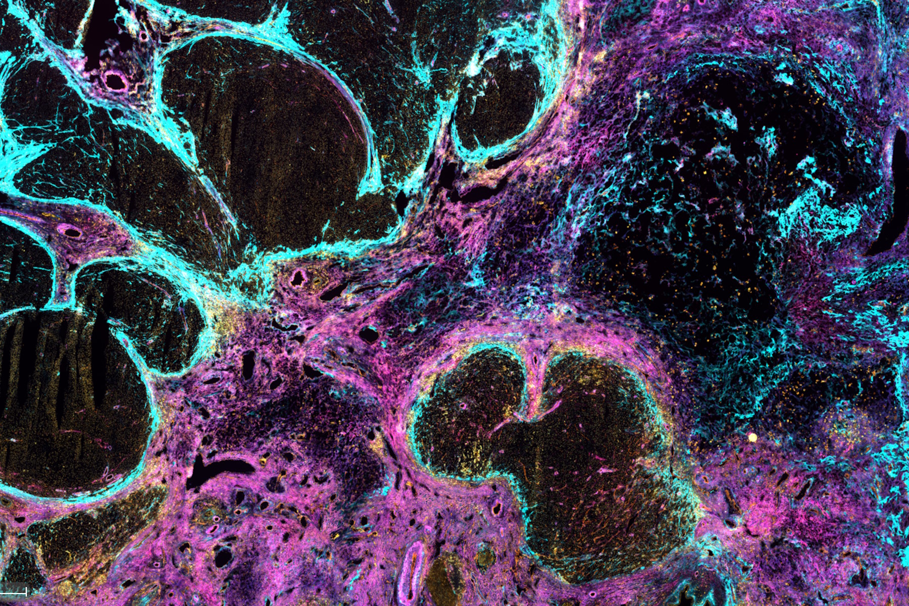
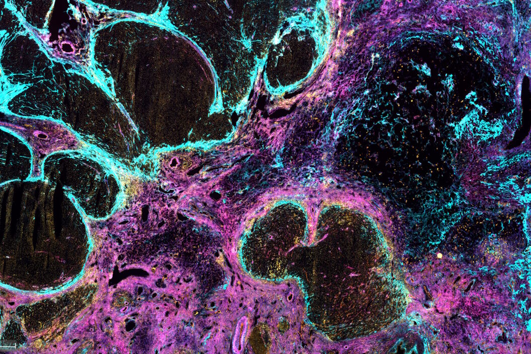
Co-detection of 10 extracellular matrix proteins and 3 topographical tissue landmarks by multiplex immunostaining within a single high-grade fibrous hotspot from a human hepatocellular carcinoma Single_high-grade_fibrous_hotspot_from_a_human_hepatocellular_carcinoma.jpg

Here, we explore a groundbreaking multiplex immunodetection approach to identifying cancer stem cell niches within hepatocellular carcinoma (HCC), focusing on the in-situ localization of extracellular matrix (ECM) signatures via multiplex imaging. HCC, a major cause of cancer-related deaths, often emerges in patients with chronic fibroinflammatory liver diseases. These diseases lead to ECM protein complex deposition, altering tissue architecture and promoting HCC development. The ECM, comprising various proteins acts as both a scaffold and a signaling hub for cellular functions, so its detection is vital in advancing our understanding of this disease. Read on to find out how Cell DIVE can help identify intratumoral structures using proteomics.

About the Application Note

Key Learnings 

  • How intratumoral structures are identified using proteomics.
  • Identifying toppgraphcial tissue landmarks and extracellular matrix proteins.
  • A guide to multiplexed immunohistochemical analyses with Cell DIVE.
  • Identification, labelling and analysis of antibodies from immunostained tissue sample images.

Learn about the transformative potential of multiplexed immunodetection

Multiplexed imaging plays a crucial role in advancing cancer research by allowing scientists to delve deep into the complex microenvironments of tumors, unveiling unique characteristics and dynamic behaviors. This revolutionary technique concurrently visualizes various cell populations and their interactions within the tumor microenvironment, providing profound insights into the intricate ecosystem of tumors. This is indispensable for interpreting the tumor's reaction to therapeutic interventions, predicting treatment outcomes, and pinpointing potential targets for immunotherapeutic strategies. Multiplexed imaging is a vital tool aiding the ongoing quest to uncover cancer stem cell niches in hepatocellular carcinoma.

This application note underscores the transformative potential of multiplexed immunodetection in identifying and characterizing cancer stem cell niches within hepatocellular carcinoma (HCC). By simultaneously detecting a diverse array of extracellular matrix (ECM) proteins and tissue landmarks, this innovative approach reveals the intricate molecular composition of fibrous nests. These specialized tumor microenvironments have significant implications for the understanding of disease progression and overcoming treatment resistance.

The integration of proteomics and multiplexed imaging empowers researchers to uncover the intricate nuances of cancer stem cell niches in hepatocellular carcinoma. The capabilities of Cell DIVE technology, coupled with the ClickWell slide carrier, are shown to streamline the process of multiplex immunostaining and reveal insights into intratumoral structures, fibrous nests and ECM. With comprehensive antibody validation and an iterative staining approach facilitated by Cell DIVE, researchers can simultaneously detect numerous proteins, enabling a deeper understanding of the tumor microenvironment.
Download the Application Note!

By clicking on the SUBMIT button, I confirm that I have reviewed and agree with Leica Microsystems GmbH Terms of Use and their Privacy Policy. I also understand my privacy choices as they pertain to my personal data, as detailed in the aforementioned Privacy Policy under ‘’Your Privacy Choices’’.

Related Articles

Background image
Scroll to top