 tissue on a single slide.")
About the case study
Key Learnings
- Learn how CST validates antibodies for Cell DIVE
- Understand the range of Cell DIVE-validated antibodies from CST
- See the antibodies in action on a wide range of cancer and tumor cell lineages
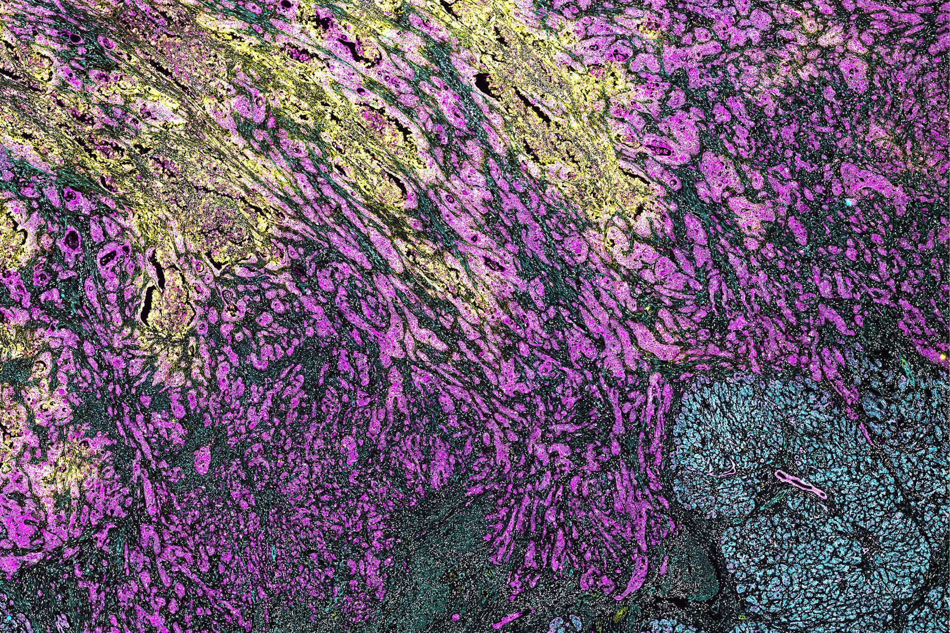
It is crucial to understand how the tumor microenvironment (TME) evolves during tumorigenesis and therapeutic response to developing personalized treatments. One of the goals is to find ways in improving cancer therapy. Immune biomarker antibodies can be used to interrogate immune cell lineages and structures with robust multiplexed imaging technologies. This approach can capture the immune response within the TME in a variety of neoplasms when is combined with specific oncology biomarker antibodies. The Cell DIVE™ Multiplex Imaging Solution allows probing and imaging of dozens of biomarkers on a whole single tissue section while using an iterative staining and dye inactivation workflow. The broad portfolio of robust IHC-validated antibodies from CST enables the detection of key proteins in the TME.
In this study, we demonstrate multiplexed Cell DIVE imaging using a novel CST panel to probe pancreatic ductal adenocarcinoma (PDAC). These biomarker antibodies define the immune cell landscape in the hypoxic tumor. CST antibodies undergo a vigorous validation process to ensure antibody performance on FFPE tissue. All antibodies in this study were direct conjugates or off the shelf. The development of antibody panel required minimal optimization. It enabled the identification of complex cell types and revealed their cell-to-cell interactions within the tumor microenvironment. The availability of specific biomarkers to interrogate cell types with the use of multiplexed tissue imaging provides unprecedented novel insights. This also provides spatial resolution of immune cell populations with many cell types in the TME.
The understanding of tumor microenvironment is essential in cancer research. It can help to unveil new mechanisms that contribute to poor patient outcomes. The tumor microenvironment is complex. It is commonly heterogenous both within a single sample, as well as, across patient samples. The broad portfolio of robust IHC-validated antibodies from CST enables the detection of key proteins in the TME. Furthermore, the iterative multiplexed staining and imaging enable TME interrogation across tissue samples even in cases where tissue availability is limited.
")


