
Introduction & Aim
Pancreatic tumors are highly heterogeneous, often with more aggressive regions that are responsible for invasion and metastasis. These aggressive pancreatic ductal (PDAC) cells undergo epithelial to mesenchymal transition and create subregions in the tumor that evade treatments and provide a critical support niche for continued tumor growth and metastasis. Conventional chemotherapy plus radiation, or in advanced disease chemotherapy plus targeted drug therapy can lengthen patient survival. However, even in patients with local disease, the 5-year survival rate remains at around 40%, demonstrating that there is need for improved conventional and immune therapies for all pancreatic cancer patients. In this study, dozens of biomarkers have been used to probe tumor heterogeneity in pancreatic ductal adenocarcinoma (PDAC) tissue. We rely on the published finding that hypoxia gene expression is consistent in PDAC, regardless of tumor location, whether primary tumor or metastasis. With the spatial biology solution, Cell DIVE and proprietary Spatial AI analytics software, dozens of biomarker can be used to computationally reduce tumor heterogeneity, and to spatially define cells in the microenvironment within hypoxic and normoxic regions of PDAC, and in normal pancreas. We found that these hypoxic PDAC regions are devoid of immune cells in general, with the appearance of spatially co-localized populations of cell types consistent with tumor desmoplasis and inflammation. Taken together, multiplexed whole slide imaging and analysis enable spatially resolved whole tissue analysis of the tumor microenvironment, including new insights into spatial biology in the hypoxic PDAC environment.
Methods & Materials
For this study, an FFPE sample of Pancreatic Ductal Adenocarcinoma was obtained from a commercial source (Pantomics). Antibodies undergo a vigorous standardized validation process (Gerdes et al., 2013) [1] to ensure that antibody conjugate performance is similar to primary secondary staining on tissues. Thirty biomarkers from various vendors were used in this study, as supplied commercial conjugates or following in-house conjugation. Slides were iteratively stained using the Leica Bond (Leica Biosystems). Slides were imaged on the Cell DIVE using four channels plus DAPI, with automatic AF removal, corrections and stitching. Fully stitched images were imported, fused, visualized, and analyzed using proprietary software developed by Leica Microsystem.
Results
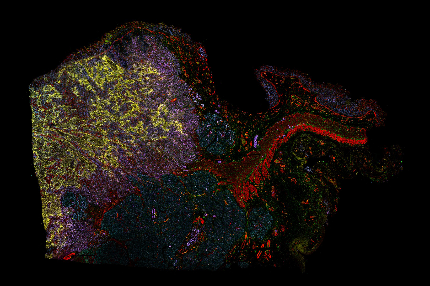
Using Cell DIVE multiplexed imaging solution, a PDAC FFPE section was iteratively stained with 30 biomarkers to spatially define aggressive tumor cells. The markers used in this study define metabolic and hypoxic, apoptotic, EMT, and immune cell status spatially within the tumor (Figure 1A). Specifically, the hypoxia marker Glut1, and two PanCK biomarkers separate cancer cells with increased glucose uptake, a cancer hallmark (Figure 1B). Following segmentation using the full set of 30 biomarkers, a small subset of biomarkers was used to classify and spatially characterize heterogeneous regions of the tumor (Figure 1C). Analyzing data in this way reduces heterogeneity by analyzing cell types in more aggressive regions of the tumor separately from cells in less aggressive regions or in normal tissue. Quantifying immune contribution across regions, normoxic tumor and normal pancreas compared to hypoxic tumor regions, there is an overll reduction in lymphocytes, and increased CD45+ populations, even though the total cells in all regions are similar (Figure 1D). Representative phenotype images are shown (Figure 1D bottom panel). Then clustering further defines the cell populations without bias.

Cluster analysis and dimension reduction revealed fewer populations in the hypoxic tumor region compared to the other regions (Figure 2). Similar to immune cell counts based on phenotyping, very few lymphocytes are present in the hypoxic tumor regions (Figure 2C Cluster and UMAP). Interestingly, several clusters reveal populations of cancer relationships clearly define cell types commonly found in the same spatial neighborhood as the CAF’s (Figure 2C; Neighborhood charts). For example, Cluster 3 (SMA+ VIM+ Collagen+ CAF cluster) and Cluster 7 (Immune Cell: CD45+, Collagen+) are common neighbors, indicative of inflammation and tumor desmoplasia commonly associated with poor survival outcomes in PDAC patients [2].

Conclusions
Our study demonstrated iterative staining and imaging of a single PDAC section with 30 biomarkers using the Cell DIVE multiplexed imaging solution. This combination of multiplexed imaging and single cell analysis within the tumor microenvironment is powerful and enables a detailed examination of single cell biomarker expression in the context of the aggressive region of a tumor. Here, the contribution of the extracellular matrix, inflammatory cells, and cancer associated fibroblasts have been spatially defined within the TME. Future work will be to further define the ECM, CAF, and inflammatory components in PDAC. For example, beyond C0L1A1, other collagens, fibronectin and SPARC can be explored, as well as additional factors involved in CAF infiltration and inflammation. Importantly, Cell DIVE multiplexing solution is tissue preserving, and biomarker expression can continue to be explored on the same tissue section and overlayed with all previously stained biomarkers from the study. Characterizing the tumor microenvironment in aggressive regions of the tumor can help to elucidate new mechanisms that contribute to poor patient outcomes and may lead to the discovery of novel interventions to improve future therapeutic success in the treatment of PDAC.
