
Cell DIVE multiplexed immunodetection in cancer research
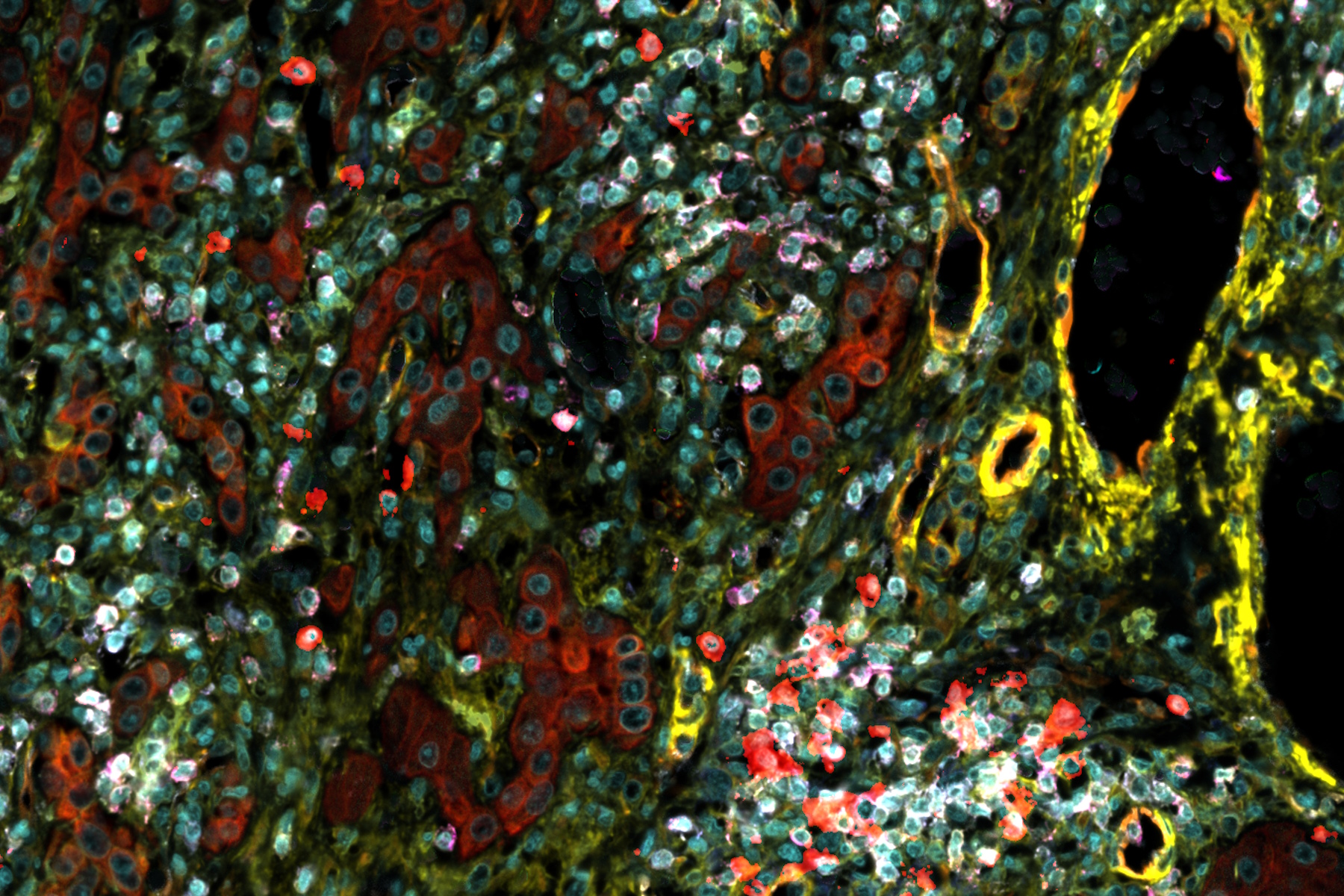
Explore the transformative potential of multiplexed immunodetection in cancer research, as demonstrated by Cell DIVE. Understand how this technology empowers researchers to uncover intricate details of cancer cellular heterogeneity via protein marker multiplex imaging. Dr. Cheung showcases the integration of proteomics and multiplexed imaging, providing a comprehensive guide to identifying the cellular expression of various receptors and their spatial relationship in the tumor microenvironment.
Understanding tumor heterogeneity with protein marker expression patterns
Dr. Cheung's research using Cell DIVE technology unveils the complexities of tumor heterogeneity, offering a guide to understanding the variations in protein marker expression patterns within cancer epithelium. Witness the spatial arrangement of key markers such as Estrogen Receptor, Progesterone Receptor, HER2, and more in different tumor subtypes. This session provides critical insights for precision medicine, aiding in the identification of potential targets and personalized treatment strategies based on the unique molecular signatures of individual tumors.
Practical insights with Cell DIVE: Multiplexed imaging in cancer analysis
Gain practical insights into multiplexed immunohistochemical analyses with Cell DIVE, enabling the identification, labeling, and analysis of antibodies from immunostained tissue sample images. This revolutionary technique concurrently visualizes various cell populations within the tumor microenvironment, offering profound insights crucial for interpreting treatment responses and predicting outcomes. Join Dr. Alison Cheung as she explores the indispensable role of multiplexed imaging in advancing our understanding of tumor ecosystems, providing essential information for therapeutic interventions.


.")
