Related Articles
-
")
How to Streamline High-Plex Imaging for 3D Spatial Omics Advances
In this webinar, Dr. Julia Roberti and Dr. Luis Alvarez from Leica Microsystems introduce…
Jun 24, 2025Read article -

Transforming Research with Spatial Proteomics Workflows
Spatial Proteomics, Nature Methods 2024 Method of the Year, is driving research advancements in…
Jun 10, 2025Read article -

Get to Insights Faster and Easier with AI Image Analysis Tools
Discover how Aivia helps scientists streamline image analysis with fast setup, accurate AI…
May 22, 2025Read article
Related Pages
-

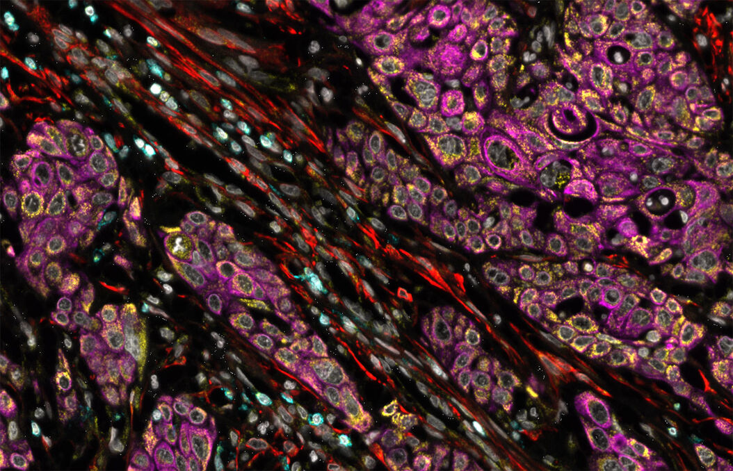
Hyperplex Cancer Tissue Analysis at Single Cell Level with Cell DIVE
The ability to study how lymphoma cell heterogeneity is influenced by the cells’ response to their…
Visit related page
