Discover how Multiplexed Bioimaging can Advance Cancer Research
Watch the Spatial Biology Symposium to explore how advanced imaging techniques can help to gain insights into cellular tumor structure and behavior

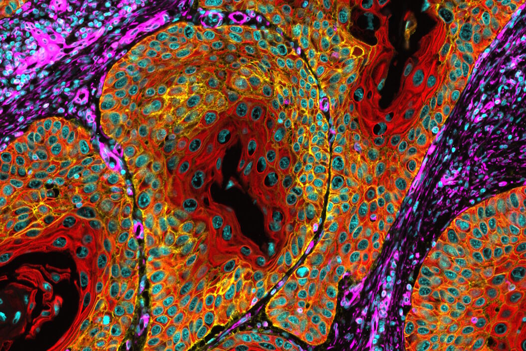
Watch an informative discussion with industry and academic experts who share their knowledge in using multiplexed imaging in research. Discover how multiplexing is revolutionizing oncology, neurology, and immunology by uncovering molecular insights that were previously elusive. Get a deeper understanding of the tissue microenvironment using advanced imaging techniques that can shed new insights into diseases such as metabolic disorders and cancer.
About the Symposium
Here are the highlights of the discussion:
- How multiplexed imaging can improve tumor treatment outcomes by revealing insights into the tumor microenvironment;
- Understand the workflow of multiplexed imaging and various methods of iterative staining;
- Uncover the potential of multimodal tissue imaging to yield high-performance biomarkers;
- How the laser-microdissection technique helps researchers to understand the underlying mechanisms of tumor growth and progression;
- Technical implementations of Cell DIVE to optimize data quality, reproducibility, and throughput.
During the symposium, a panel of four experts shared their insights on using various techniques to identify and track multiple biomarkers. They discussed the latest advancements in this field and how these techniques can improve medical research and diagnosis. Dr. Harikesh Wong talked about the imaging control of the immune response. He emphasized the importance of quantitative fluorescence microscopy methods, computational modeling, and experimental perturbations in studying intercellular circuits that trigger immune response and their emergent behaviors within intact tissue environments. Lisa Arvidson spoke about the importance of choosing reliable antibody conjugates in highly multiplexed single-cell analysis and imaging.
Dr. Jia-Ren Lin discussed the whole-specimen imaging strategy, that highlights large-scale morphological and molecular gradients, which can expand our understanding of cancer beyond discrete genetic changes. Complimenting the discussion, Doug Bowman provided key insights on analyzing highly multiplexed tissue imaging. At the end, Rick Heil-Chapdelaine provided an overview on open multiplexing using Cell DIVE that lets the user’s research dictate the level of automation required, which antibodies to use, how to build your antibody panel, and more.


