Probing Human Alzheimer's Cortical Section using Spatial Multiplexing
Enhancing our understanding of the spatial heterogeneity in human Alzheimer’s cortical section by combining multiplexed imaging and AI-guided image analysis

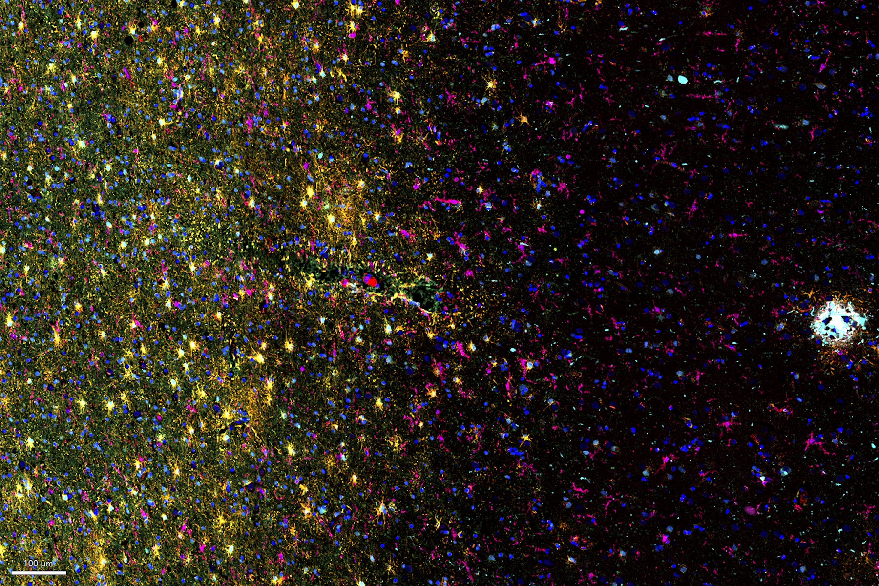
Alzheimer’s disease (AD) is the most common neurodegenerative disease and is characterized by the progressive decline of cognitive function. Spatial profiling of AD brain may reveal cellular relationships, facilitating a better understanding of disease etiology. This study captures a global overview of the AD cortical tissue composition and emphasizes the streamlined workflow of Cell DIVE imaging, from data acquisition to AI-based analysis using Aivia software, resulting in quicker insights.
Key learnings:
- Investigate the spatial distribution of AD-associated markers (e.g., the aggregation patterns of Tau proteins and β -amyloid plaques) throughout the cortical tissue of the brain.
- Characterize the neuronal loss by identifying cell types affected by neurodegeneration in AD cortical tissue.
- Explore the spatial landscape of neuroinflammation by visualizing microglia and astrocytes relative to the B-amyloid plaques.
- Discover the benefits of transforming tissue research with the Cell DIVE multiplex imaging solution and AI-powered image analysis using Aivia.
In this study, we demonstrate multiplexed Cell DIVE imaging using a novel CST® panel to probe AD cortical tissue section. Further, using AI-guided analysis with AIVIA provides the researcher the power to-characterize individual neuronal components and identify spatially co-localized populations of neuronal cell types with respect to β -amyloid plaques using clustering and relational analysis.


