Gold EM grid with a silicon dioxide (SiO₂) support film, containing vitrified T47D cells intrinsically expressing GFP-H2B (labeling nuclei) and RFP-LifeAct (labeling the actin cytoskeleton) and high-pressure frozen C. elegans germline tissue, fluorescently labeled with GFP-tagged synaptonemal complex protein

Flux de préparation des échantillons EM & Utilisations

Les solutions de préparation d'échantillons Leica permettent aux chercheurs d'obtenir des résultats de haute qualité, précis et reproductibles lors de l'imagerie d'échantillons en microscopie électronique. Nos solutions prennent en charge la microscopie électronique à balayage (MEB), la microscopie électronique à transmission (MET) et la cryomicroscopie électronique.

Prenez simplement contact avec nous !

Nos experts en imagerie sont là pour vous aider en vous conseillant sur les solutions pour les flux de travail de préparation des échantillons EM & Uses.

Comment les chercheurs peuvent-ils préparer les échantillons pour le MEB ?

La préparation des échantillons pour la microscopie électronique à balayage (MEB) implique des techniques telles que le revêtement, le séchage et l'enrobage afin de garantir une imagerie optimale. En utilisant les solutions Leica pour les échantillons MEB, les chercheurs améliorent la conductivité, réduisent les artefacts et garantissent des surfaces stables et de haute qualité pour une imagerie MEB fiable à température ambiante.

Quelles sont les meilleures méthodes de préparation des échantillons TEM ?

La préparation des échantillons TEM (microscopie électronique à transmission) nécessite des coupes ultrafines et sans dommages, un revêtement par pulvérisation cathodique des grilles EM et une coloration pour obtenir une visualisation détaillée de la structure. Grâce à la précision et à la fiabilité des instruments Leica, les chercheurs peuvent produire des images TEM de haute résolution.

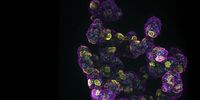
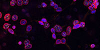
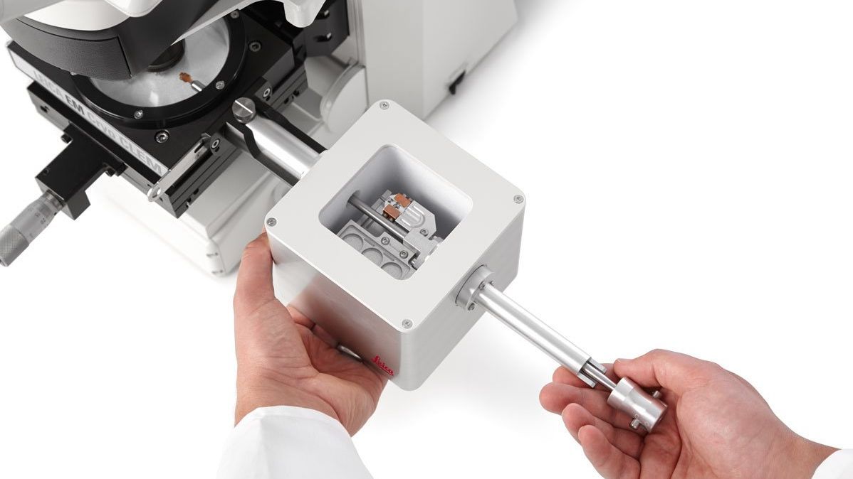
Qu'est-ce qui est important pour la préparation des échantillons de cryo-EM ?

Le Cryo EM bénéficie de la vitrification par congélation rapide pour maintenir les structures dans leur état naturel.

Les solutions Leica de préparation cryogénique - y compris la vitrification avancée, l'enrobage, la coupe ultramince, le cryoplaning et les flux de travail personnalisables de transfert cryogénique et de CLEM - fournissent des échantillons reproductibles et exempts de contamination. Ils aident à maintenir les structures natives, permettent un ciblage précis et une imagerie cryo EM à haute résolution.

Pourquoi choisir les instruments et solutions Leica pour la préparation d'échantillons EM ?


Préparation de l'échantillon pour le MEB

Les outils de préparation du MEB de Leica Microsystems comprennent des appareils de revêtement par pulvérisation cathodique sous vide poussé et des sécheurs à point critique. Ces solutions garantissent une imagerie de surface de haute qualité. De plus, les instruments Leica fournissent des résultats cohérents et fiables pour vos études MEB.


Préparation de l'échantillon TEM

Les chercheurs peuvent compter sur les solutions de préparation TEM de Leica Microsystems pour leur précision et leur fiabilité. Des outils essentiels tels que les ultramicrotomes et les systèmes de coloration automatisés permettent de produire des coupes ultrafines et des images à haute résolution. Le revêtement par pulvérisation cathodique améliore la conductivité de l'échantillon et la qualité de l'imagerie.


Préparation Cryo-EM complète

Avec les instruments de cryopréparation de Leica Microsystems, notamment les congélateurs à plongeur et les congélateurs à haute pression, les chercheurs garantissent une congélation et une vitrification rapides. En outre, notre système de transfert d'échantillons pour les flux de travail cryogéniques offre des options de transfert pour tous les types d'échantillons. Cette option les protège de la contamination, garantissant ainsi une analyse Cryo-EM précise et détaillée.

Articles connexes

Lire nos derniers articles sur Flux de préparation des échantillons EM & Utilisations

Le portail de connaissances de Leica Microsystems propose des ressources pour la recherche scientifique et l’enseignement dans le domaine de la microscopie. Le contenu a été élaboré pour aider les utilisateurs novices et expérimentés, ainsi que les scientifiques dans leurs tâches et expériences quotidiennes.

Plus d’articles
Sectioning resin-embedded sambles

Ultramicrotomy eBook: Targeting, Trimming & Alignment

Ultramicrotomy is evolving rapidly, and today’s microscopes demand high‑quality sections, precise targeting, and reproducible workflows. This eBook brings together expert application notes, automated…
Foraminifera (Ammonia confertitesta) 
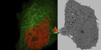
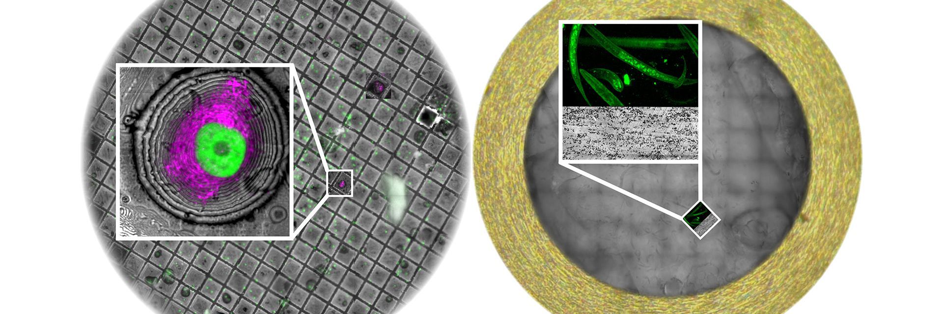
labeled with membrane-permeable calcein, high-pressure frozen in salt water using EM ICE. The sample was cryo-planed and targeted with the M205 on the Cryo-Fluo Enuity, then transferred under cryo conditions to the Cryo-Stellaris for widefield and confocal imaging, revealing details of the staining pattern. Image courtesy: David Evans, University of Southampton.

High-Pressure Freezing for Organoids: Cryo CLEM & FIB Lift Out

Master cryo EM workflow steps for challenging 3D samples: when to choose HPF vs. plunge freezing, reproducible blotting/ice control, contamination aware transfers, Cryo CLEM 3D targeting in organoids,…
Electron microscope (EM) 
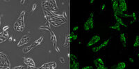
image of a cross section of C. elegans (roundworm). Courtesy of T. Müller-Reichert, MPI-CBG, Dresden, Germany and K. McDonald, University of California, Berkeley, USA.

Brief Introduction to High-Pressure Freezing for Cryo-Fixation

Preparation of biological specimens for electron microscopy (EM) often requires cryo-fixation which does not introduce significant structural alterations of cellular constituents. A common method used…
TEM micrographs of polymer sections. Left: Poly(styrene)-
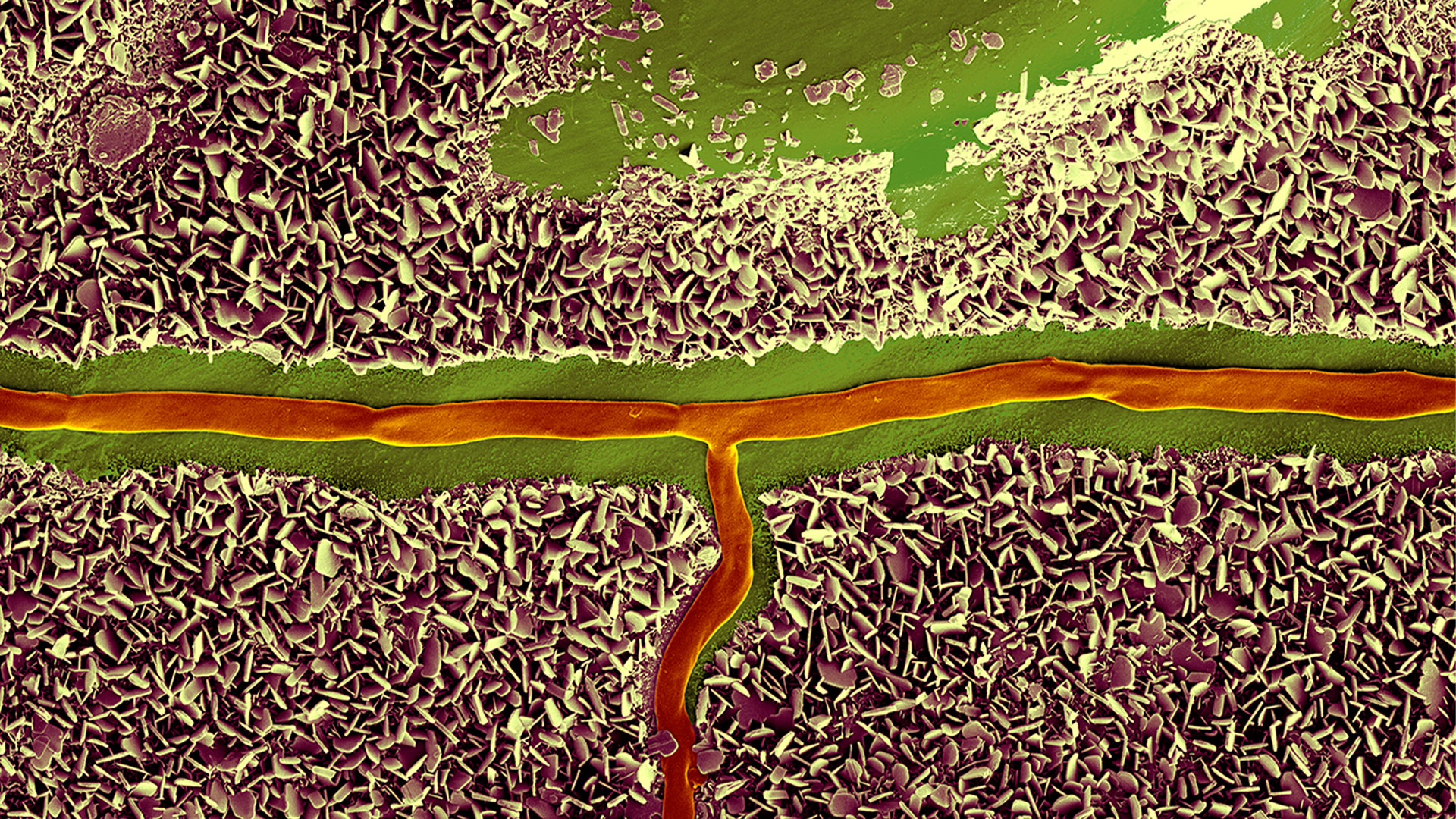
b-poly(isoprene). Right: Poly(styrene)-b-poly(methyl methacrylate).

Ultramicrotome Sectioning of Polymers for TEM Analysis

We demonstrate the capabilities of the UC Enuity ultramicrotome from Leica Microsystems for preparing ultrathin sections of polymer samples under both ambient and cryogenic conditions. By presenting…
Final Segmentation of organelles in Trichomonas species. Magenta – costa, light blue – hydrogenosomes, turquoise – ER, red – vacuoles, yellow – axostyle, green – Golgi apparatus.  Sample courtesy of Isabelle Guerin-Bonne, Low Kay En, Electron Microscopy Unit, Yong Loo Lin School of Medicine, National University of Singapore. Scale bar: 1 µm.

Volume EM and AI Image Analysis

The article outlines a detailed workflow for studying biological tissues in three dimensions using volume-scanning electron microscopy (volume-SEM) combined with AI-assisted image analysis. The focus…

Integrated Serial Sectioning and Cryo-EM Workflows for 3D Biological Imaging

This on-demand webinar explores how integrated tools can support electron microscopy workflows from sample preparation to image analysis. Experts Andreia Pinto, Adrian Boey, and Hoyin Lai present the…
SEM image of the full Li-NMC electrode sample, showing the two porous layers and the metal film at the center of the structure.

Revealing Sodium Battery Degradation via Cryo-EM and CryoFIB

Explore how cryogenic electron microscopy and focused ion beam techniques uncover the intrinsic structure of sodium battery interfaces. This webinar presents a new degradation model based on separator…
Mouse hippocampus brain slice on a grid after HPF using the “Waffle Method”.

The “Waffle Method”: High-Pressure Freeze Complex Samples

This article describes the advantages of a special high pressure freezing method, the so-called “Waffle Method”. Learn how the “Waffle Method” uses EM grids as spacers for high-pressure freezing,…
UC Enuity with Diatome diamond knife.

Mastering Polymer Sectioning with Helmut Gnaegi

When it comes to ultramicrotomy, few names carry the weight of Helmut Gnaegi. As co-founder of Diatome, a global leader in diamond knife technology, Helmut has spent decades refining the art and…
C. elegans embedded in Lowicryl® HM20; pharynx showing red fluorescence (mCherry). The overview shows a front view onto the resin capsule formed by the bottom of a flow-through chamber of the EM AFS2. The capsule was pretrimmed manually. The blockface was trimmed automatically using the AutoTrim function of UC Enuity guided by fluorescence of the worm. Edge length of both squares in relation to the images is 250 µm.

How Fluorescence Guides Sectioning of Resin-embedded EM Samples

Electron microscopes, including transmission electron microscopes (TEM) and scanning electron microscopes (SEM), are widely utilized to gain detailed structural information about biological samples or…
14: 7-day old 3D cell culture of MDCK cells stably transfected with; Mx1-GFP (green), SPY555-Actin (yellow) and WGA-AlexaTM 647 (magenta), and imaged using a THUNDER Imager Cell Spinning Disk. Shown are both the spinning disk image (left) and the same image after THUNDER Computational Clearing was applied (right); scale bar 50 μm.

Organoïdes et culture cellulaire en 3D

L’une des avancées récentes les plus passionnantes de la recherche en sciences de la vie est le développement de systèmes de culture cellulaire en 3D, tels que les organoïdes, les sphéroïdes ou les modèles d’organes sur puce. Une culture cellulaire en 3D est un environnement artificiel dans lequel les cellules sont capables de se développer et d’interagir avec leur environnement dans les 3 dimensions. Ces conditions sont similaires à celles qui existeraient in vivo.

En savoir plus

Image of C2C12 cells: The cells are stained with lamin B (magenta) which indicates nuclear structure, Hoechst (blue) indicating DNA, and γH2AX (yellow) indicating damage to DNA. Cells were imaged using a THUNDER Imager 3D Live Cell with a 63X/1.4 oil immersion objective.

Recherches en biologie cellulaire

L’imagerie en biologie cellulaire est réalisée à l’aide d’une gamme de microscopes photoniques et électroniques. Les solutions d’imagerie proposées par Leica Microsystems sont conçues pour maximiser vos recherches en biologie cellulaire.

En savoir plus

Mammalian cells expressing H2B-mCherry and alphaTubulin-mEGFP to visualize DNA and microtubules respectively. Left: Widefield imaging using THUNDER Imager. Right: Sample after correlated EM imaging using Leica Microsystems Coral Life workflow. Cells were cryo-immobilized by high-pressure freezing 60 min after the onset of cytokinesis. The intercellular bridge was imaged under TEM after freeze substitution and resin infiltration.

Microscopie corrélative CLEM

Les solutions CLEM de Leica Microsystems garantissent la viabilité des échantillons, les contrôles de qualité et un mécanisme de ciblage 3D précis et fiable. Les utilisateurs peuvent tirer parti de ces solutions pour identifier directement la bonne cellule au bon moment, obtenir des données cryogéniques confocales haute résolution ou placer les informations de fluorescence dans le contexte ultrastructural.

En savoir plus

Culture cellulaire

La culture de cellules en laboratoire constitue la base des travaux des scientifiques spécialisés dans les domaines de la biologie cellulaire, la recherche contre le cancer, la biologie du développement ou tout autre domaine de recherche en sciences de la vie ou dans le domaine pharmaceutique. Découvrez comment Leica contribue à la culture de cellules animales en laboratoire.

En savoir plus

14: 7-day old 3D cell culture of MDCK cells stably transfected with; Mx1-GFP (green), SPY555-Actin (yellow) and WGA-AlexaTM 647 (magenta), and imaged using a THUNDER Imager Cell Spinning Disk. Shown are both the spinning disk image (left) and the same image after THUNDER Computational Clearing was applied (right); scale bar 50 μm.
Image of C2C12 cells: The cells are stained with lamin B (magenta) which indicates nuclear structure, Hoechst (blue) indicating DNA, and γH2AX (yellow) indicating damage to DNA. Cells were imaged using a THUNDER Imager 3D Live Cell with a 63X/1.4 oil immersion objective.
Mammalian cells expressing H2B-mCherry and alphaTubulin-mEGFP to visualize DNA and microtubules respectively. Left: Widefield imaging using THUNDER Imager. Right: Sample after correlated EM imaging using Leica Microsystems Coral Life workflow. Cells were cryo-immobilized by high-pressure freezing 60 min after the onset of cytokinesis. The intercellular bridge was imaged under TEM after freeze substitution and resin infiltration.
Scroll to top