Leica DM3000 & DM3000 LED
Microscopes droits
Microscopie optique
Produits
Accueil
Leica Microsystems
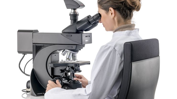
Leica DM3000 & DM3000 LED Microscopes de système particulièrement ergonomiques avec automatisation intelligente
Amélioration du flux de travail du laboratoire
Lire nos derniers articles
Factors to Consider when Selecting Clinical Microscopes
What matters if you would like to purchase a clinical microscope? Learn how to arrive at the best buying decision from our Science Lab Article.
Differential Interference Contrast (DIC) Microscopy
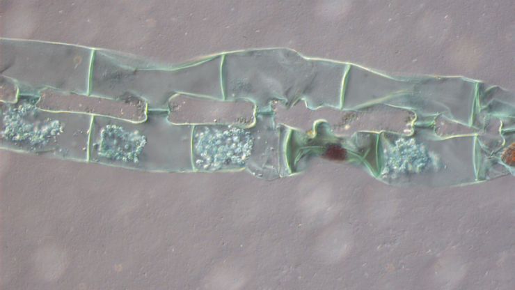
This article demonstrates how differential interference contrast (DIC) can be actually better than brightfield illumination when using microscopy to image unstained biological specimens.
H&E Staining in Microscopy
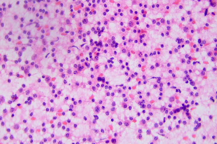
If we consider the role of microscopy in pathologists’ daily routines, we often think of the diagnosis. While microscopes indeed play a crucial role at this stage of the pathology lab workflow, they…
How to Benefit from Digital Cytopathology
If you have thought of digital cytopathology as characterized by the digitization of glass slides, this webinar with Dr. Alessandro Caputo from the University Hospital of Salerno, Italy will broaden…
The Time to Diagnosis is Crucial in Clinical Pathology
Abnormalities in tissues and fluids - that’s what pathologists are looking for when they examine specimens under the microscope. What they see and deduce from their findings is highly influential, as…
A Guide to Darkfield Microscopes
A darkfield microscope offers a way to view the structures of many types of biological specimens in greater contrast without the need of stains.
A Guide to Phase Contrast
A phase contrast light microscope offers a way to view the structures of many types of biological specimens in greater contrast without the need of stains.
Perform Microscopy Analysis for Pathology Ergonomically and Efficiently
The main performance features of a microscope which are critical for rapid, ergonomic, and precise microscopic analysis of pathology specimens are described in this article. Microscopic analysis of…
Koehler Illumination: A Brief History and a Practical Set Up in Five Easy Steps
In this article, we will look at the history of the technique of Koehler Illumination in addition to how to adjust the components in five easy steps.
Fields of Application
Pathologie clinique
Découvrez comment les solutions de microscopie de pathologie Leica aident les pathologistes cliniques à diagnostiquer les infections et les maladies à partir des fluides et des tissus corporels.
Microscopie de pathologie
L’analyse d’échantillons pour la pathologie nécessite parfois de longues heures de travail au microscope. Pour l’utilisateur, il peut en résulter un inconfort physique et une tension qui peuvent…
Pathologie anatomique
Découvrez comment les microscopes d'anatomie pathologique de Leica Microsystems permettent de réaliser des diagnostics médicaux efficaces et précis.
