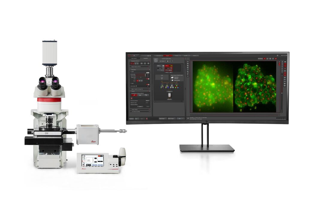
THUNDER Imager EM Cryo CLEM
THUNDER Imaging System
Prodotti
Home
Leica Microsystems
THUNDER Imager EM Cryo CLEM
Conoscenza approfondita della biologia strutturale delle cellule
Leggi gli articoli più recenti
How to Successfully Perform Live-cell CLEM
The Leica Nano workflow provides a streamlined live-cell CLEM solution for getting insight bout structural changes of cellular components over time. Besides the technical handling described in the…
How to Successfully Implement Coral Life
The live-cell CLEM workflow allows you to capture dynamic information related to a relevant biological process as it happens and put these observations into their ultrastructural context. The Leica…
Advancing Cellular Ultrastructure Research
Freeze-fracture and freeze-etching are useful tools for studying flexible membrane-associated structures such as tight junctions or the enteric glycocalyx. Freeze-fracture and etching are two…
The Cryo-CLEM Journey
This article describes the Cryo-CLEM technology and the benefits it can provide for scientists. Additionally, some scientific publications are highlighted.
Recent developments in cryo electron…
Download EM Workflow Solutions Booklet
This publication is a compilation of appropriate workflows for the most frequently used sample preparation methods, like Correlative Methodologies, Optogenetics & Electro-Physiology, Surface Analysis,…
Exploring the Structure and Life Cycle of Viruses
The SARS-CoV-2 outbreak started in late December 2019 and has since reached a global pandemic, leading to a worldwide battle against COVID-19. The ever-evolving electron microscopy methods offer a…
Targeting Active Recycling Nuclear Pore Complexes using Cryo Confocal Microscopy
In this article, how cryo light microscopy and, in particular cryo confocal microscopy, is used to improve the reliability of cryo EM workflows is described. The quality of the EM grids and samples is…
Advancing Cell Biology with Cryo-Correlative Microscopy
Correlative light and electron microscopy (CLEM) advances biological discoveries by merging different microscopes and imaging modalities to study systems in 4D. Combining fluorescence microscopy with…
Workflows and Instrumentation for Cryo-electron Microscopy
Cryo-electron microscopy is an increasingly popular modality to study the structures of macromolecular complexes and has enabled numerous new insights in cell biology. In recent years, cryo-electron…
An Introduction to Computational Clearing
Many software packages include background subtraction algorithms to enhance the contrast of features in the image by reducing background noise. The most common methods used to remove background noise…
Crystal Clear Cryo Light-microscopy Images
This article describes how computational clearing of cryo light microscopy images improves the identification of cellular targets for cryo electron-microscopy.
Real Time Images of 3D Specimens with Sharp Contrast Free of Haze
THUNDER Imagers deliver in real time images of 3D specimens with sharp contrast, free of the haze or out-of-focus blur typical of widefield systems. They can even image clearly places deep inside a…
Campi di applicazione
Microscopia a fluorescenza
La fluorescenza è uno dei fenomeni fisici più comunemente utilizzati nella microscopia biologica e analitica, soprattutto a causa della sua elevata sensibilità e specificità. Scopri come i microscopi…
Ricerca sul cancro
Il cancro è una malattia complessa ed eterogenea causata da cellule che hanno dei difetti nella regolazione della crescita. I cambiamenti genetici ed epigenetici in una cellula o in un gruppo di…
Biologia cellulare
Se la tua ricerca è incentrata sulla comprensione delle basi cellulari delle condizioni di salute e delle malattie umane, è fondamentale analizzare le cellule d’interesse con un dettaglio…
Virology
L’oggetto della tua ricerca si concentra su infezioni e malattie virali? Scopri in che modo puoi approfondire l’aspetto virologico utilizzando le soluzioni di imaging e la preparazione del campione di…
Organismi modello nella ricerca
Un organismo modello è una specie utilizzata dai ricercatori per studiare specifici processi biologici. Hanno caratteristiche genetiche simili a quelle umane e vengono normalmente impiegate in aree di…
Tomografica crioelettronica
La tomografia crioelettronica (CryoET) viene utilizzata per rilevare le biomolecole all'interno del proprio ambiente cellulare fino a una risoluzione senza precedenti, inferiore a un nanometro.
Tecniche avanzate di microscopia
Le tecniche di microscopia avanzata comprendono tecniche di imaging ad alta e super-risoluzione. Queste tecniche sono utilizzate principalmente per visualizzare gli eventi biologici ad altissima…
Microscopia correlativa per sistemi ottici ed elettronici (CLEM)
I flussi di lavoro Coral di Leica Microsystems consentono agli utenti di correlare i dati di fluorescenza e microscopia elettronica (CLEM).