電顕試料作製ワークフロー&アプリケーション

ライカの電顕試料作製ソリューションを使用することで、電子顕微鏡によるサンプルのイメージングにおいて、高品質、高精度、再現性の高い結果を安定して得ることができます。ライカのソリューションは、走査型電子顕微鏡(SEM)、透過型電子顕微鏡(TEM)、クライオ電子顕微鏡 (CryoEM)をサポートしています。

お気軽にご連絡ください!

ライカのエキスパートが、電顕試料作製ワークフローとお客様のアプリケーションに適したソリューションをご提供します。

研究者はどのようにしてSEM用の試料を準備するのか?

SEM(走査型電子顕微鏡)用の試料調製において、コーティング、乾燥、包埋などの技術を用いることで、最適なイメージングが実現します。SEMサンプルにライカのソリューションを使用することで、研究者は導電性を改善し、アーティファクトを低減し、室温での信頼性の高いSEMイメージングのための安定した高品質の試料面を保つことができます。

TEMサンプルの前処理にはどのような方法が最適ですか?

TEM(透過型電子顕微鏡)の試料作製には、超薄切片、損傷のない切片、EMグリッドのスパッタコーティング、および詳細な構造の可視化するための染色が必要になります。ライカの装置の精度と信頼性により、研究者は高解像のTEM画像を撮影することができます。

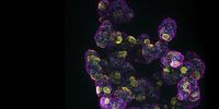
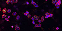
クライオ電子顕微鏡 (CryoEM)のサンプル前処理で重要なことは何ですか?

急速凍結により非晶質化することで、試料の構造を自然の状態に保つことができます。

ライカのクライオ試料作製ソリューションは、高度な非晶質化、コーティング、超薄切片化、クライオ下での断面出し、カスタマイズ可能なクライオトランスファーとCLEMワークフローを備えており、再現性が高くコンタミのない試料作製を実現します。試料本来の構造を維持することで正確なターゲティングが可能になり、高解像の Cryo EMイメージングを実現します。

電顕試料作製にライカの装置とソリューションを選ぶ理由とは?


SEMサンプルの準備

ライカマイクロシステムズのSEM試料作製ツールには、高真空スパッタコーターと臨界点乾燥装置があります。これらのソリューションにより、高品質な試料面のイメージングを可能にします。さらに、ライカの装置はSEM研究に一貫した信頼性の高い結果を提供します。


TEM試料作製

ライカマイクロシステムズのTEM試料作製ソリューションの精度と信頼性の高さを、研究者の皆さまからご評価いただいています。ウルトラミクロトームや自動染色システムのような必須ツールは、超薄切片の作製や高解像画像の撮影に役立ちます。スパッタコーティングは、試料の導電性と画像品質を向上させます。


包括的なクライオ電子顕微鏡試料作製

浸漬凍結装置や高圧凍結装置など、ライカマイクロシステムズのクライオ試料作製装置により、迅速に非晶質化での凍結を実現します。さらに、クライオワークフロー用のサンプルトランスファーシステムは、あらゆる種類のサンプルに対応するトランスファーオプションを提供します。このオプションは、コンタミネーションから試料を保護し、正確で詳細なクライオ電子顕微鏡解析を可能にします。

電顕試料作製ワークフローとアプリケーションに関するよくある質問

Show answer 創薬や医薬品開発において、なぜ生死判別アッセイを活用するのでしょうか?

患者の安全が最優先です。従って、致死量に関する知識は非常に重要であり、臨床試験を検討する前に規制当局から要求されるのです。生死判別アッセイを用いることで、閾値を評価することが可能となります。十分な量のデータを効率的に取得するために、Micaは最適なソリューションです。

Show answer レーザーマイクロダイセクションによって、バイオ医薬品研究はどのように改善されるのでしょうか?

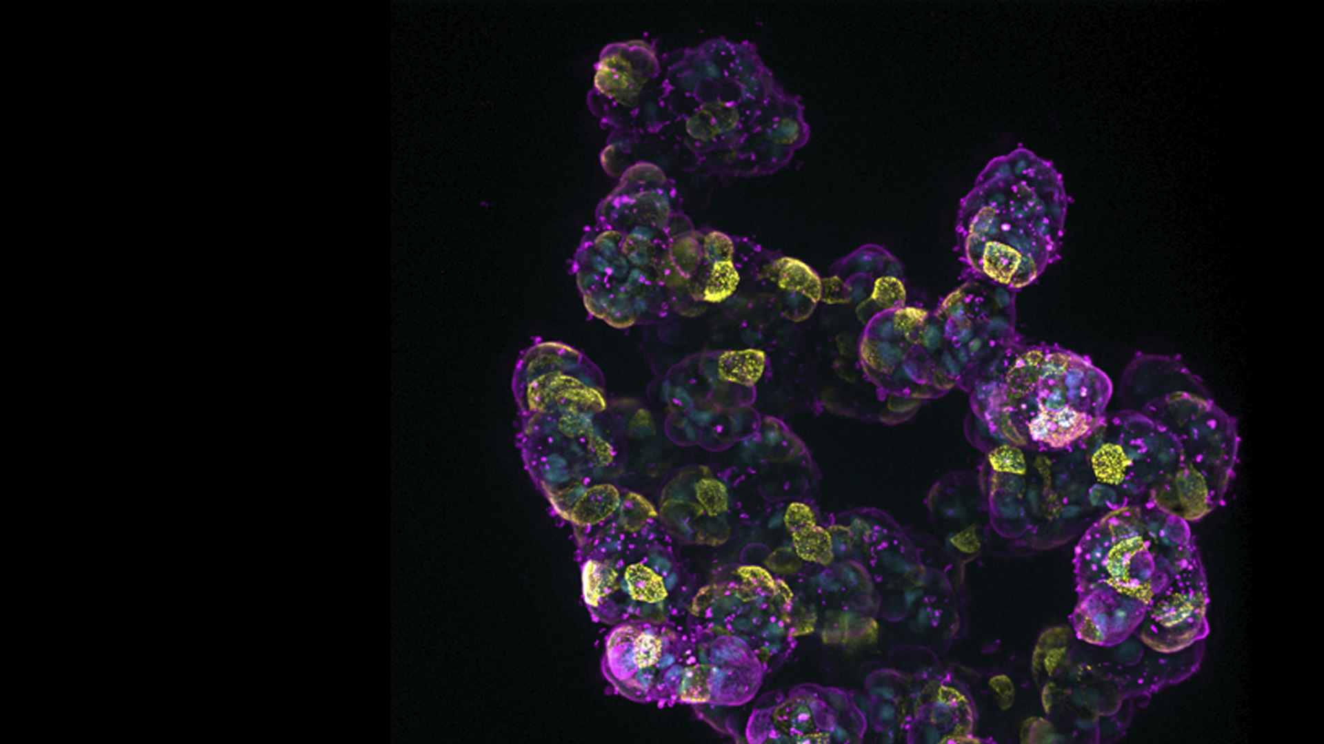
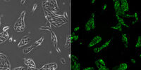
高精度に組織領域を回収することで、高品質のゲノムおよびプロテオーム解析を可能にします。レーザーマイクロダイセクションは、バイオ医薬品研究者が特定の細胞や組織領域をターゲットとし、医薬品開発においてより正確でコンタミネーションのない研究を促進するのに役立ちます。

Show answer 創薬や医薬品開発に関するバイオファーマの研究になぜ顕微鏡を使うのでしょうか?

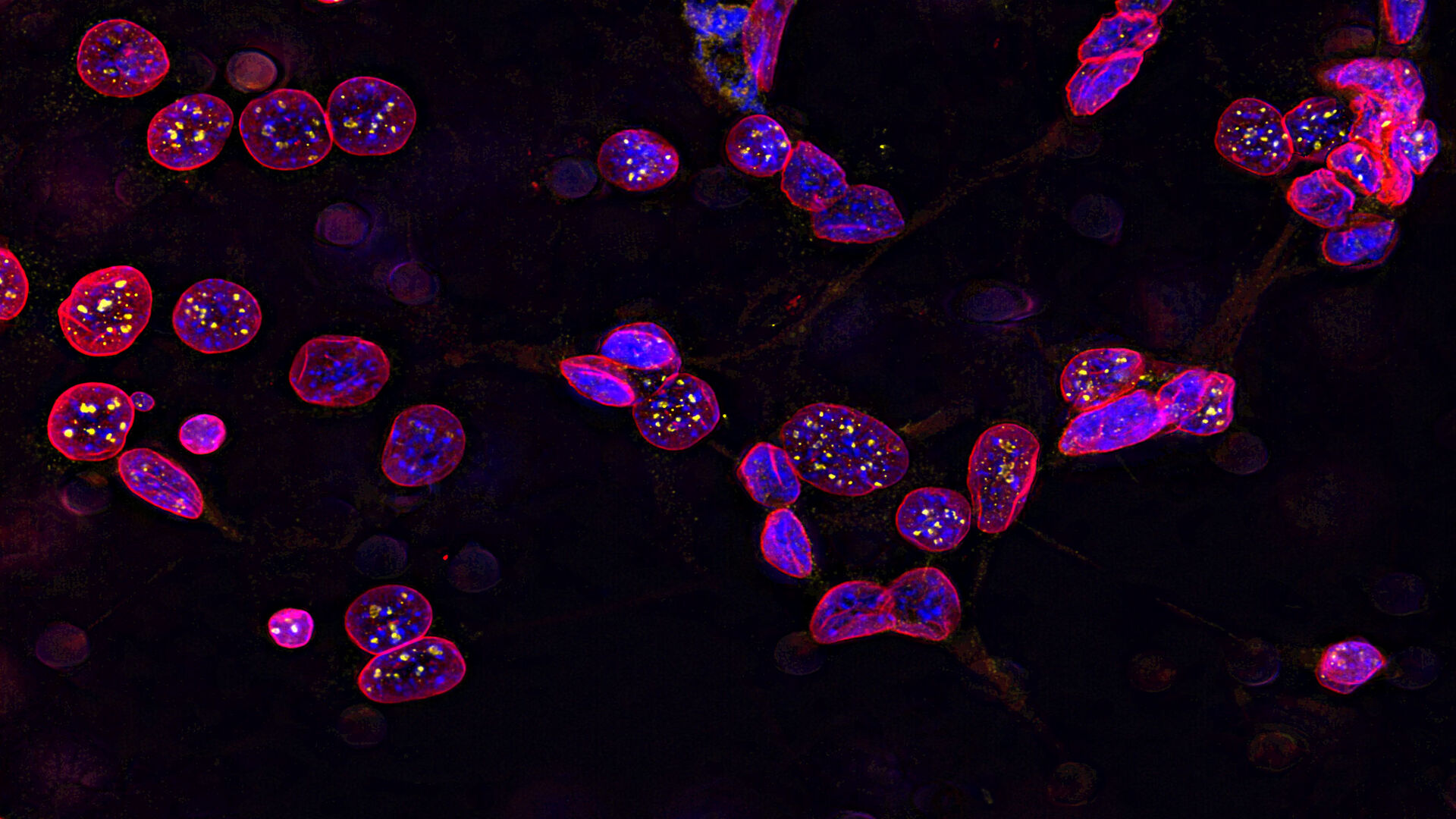
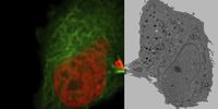
顕微鏡観察は、化合物や薬剤候補に対する細胞応答の詳細な空間的知見を得るのに役立ちます。創薬段階、すなわち生細胞培養モデルや組織の品質管理や反応経路の詳細な分析には、さまざまな創薬・開発ニーズに対応するライカ製品があります。

関連記事

電顕試料作製ワークフローに関する最新記事を読む

ライカ マイクロシステムズの知識ポータルでは、顕微鏡に関する科学的な研究記事および教材を提供しています。日常業務や研究で、ビギナーの方から経験豊富な方まで幅広くサポートします。

その他の記事
Claudia Mayrhofer ZFE Graz

Ultramicrotome UC Enuity in Practice: Stable 15 nm Sections at ZFE

After using the UCT and UC6 ultramicrotomes, Claudia Mayrhofer calls UC Enuity a leap in stability—so robust that vibrations and temperature shifts don’t spoil sections, even with multiple users. Auto…
Sectioning resin-embedded sambles

Ultramicrotomy eBook: Targeting, Trimming & Alignment

Ultramicrotomy is evolving rapidly, and today’s microscopes demand high‑quality sections, precise targeting, and reproducible workflows. This eBook brings together expert application notes, automated…
Foraminifera (Ammonia confertitesta) labeled with membrane-permeable calcein, high-pressure frozen in salt water using EM ICE. The sample was cryo-planed and targeted with the M205 on the Cryo-Fluo Enuity, then transferred under cryo conditions to the Cryo-Stellaris for widefield and confocal imaging, revealing details of the staining pattern. Image courtesy: David Evans, University of Southampton.

High-Pressure Freezing for Organoids: Cryo CLEM & FIB Lift Out

Master cryo EM workflow steps for challenging 3D samples: when to choose HPF vs. plunge freezing, reproducible blotting/ice control, contamination aware transfers, Cryo CLEM 3D targeting in organoids,…
Electron microscope (EM) image of a cross section of C. elegans (roundworm). Courtesy of T. Müller-Reichert, MPI-CBG, Dresden, Germany and K. McDonald, University of California, Berkeley, USA.

Brief Introduction to High-Pressure Freezing for Cryo-Fixation

Preparation of biological specimens for electron microscopy (EM) often requires cryo-fixation which does not introduce significant structural alterations of cellular constituents. A common method used…
TEM micrographs of polymer sections. Left: Poly(styrene)-b-poly(isoprene). Right: Poly(styrene)-b-poly(methyl methacrylate).

Ultramicrotome Sectioning of Polymers for TEM Analysis

We demonstrate the capabilities of the UC Enuity ultramicrotome from Leica Microsystems for preparing ultrathin sections of polymer samples under both ambient and cryogenic conditions. By presenting…
Final Segmentation of organelles in Trichomonas species. Magenta – costa, light blue – hydrogenosomes, turquoise – ER, red – vacuoles, yellow – axostyle, green – Golgi apparatus.  Sample courtesy of Isabelle Guerin-Bonne, Low Kay En, Electron Microscopy Unit, Yong Loo Lin School of Medicine, National University of Singapore. Scale bar: 1 µm.

Volume EM and AI Image Analysis

The article outlines a detailed workflow for studying biological tissues in three dimensions using volume-scanning electron microscopy (volume-SEM) combined with AI-assisted image analysis. The focus…

Integrated Serial Sectioning and Cryo-EM Workflows for 3D Biological Imaging

This on-demand webinar explores how integrated tools can support electron microscopy workflows from sample preparation to image analysis. Experts Andreia Pinto, Adrian Boey, and Hoyin Lai present the…
SEM image of the full Li-NMC electrode sample, showing the two porous layers and the metal film at the center of the structure.

Revealing Sodium Battery Degradation via Cryo-EM and CryoFIB

Explore how cryogenic electron microscopy and focused ion beam techniques uncover the intrinsic structure of sodium battery interfaces. This webinar presents a new degradation model based on separator…
Mouse hippocampus brain slice on a grid after HPF using the “Waffle Method”.

The “Waffle Method”: High-Pressure Freeze Complex Samples

This article describes the advantages of a special high pressure freezing method, the so-called “Waffle Method”. Learn how the “Waffle Method” uses EM grids as spacers for high-pressure freezing,…
UC Enuity with Diatome diamond knife.

Mastering Polymer Sectioning with Helmut Gnaegi

When it comes to ultramicrotomy, few names carry the weight of Helmut Gnaegi. As co-founder of Diatome, a global leader in diamond knife technology, Helmut has spent decades refining the art and…
14: 7-day old 3D cell culture of MDCK cells stably transfected with; Mx1-GFP (green), SPY555-Actin (yellow) and WGA-AlexaTM 647 (magenta), and imaged using a THUNDER Imager Cell Spinning Disk. Shown are both the spinning disk image (left) and the same image after THUNDER Computational Clearing was applied (right); scale bar 50 μm.

オルガノイドと3D細胞培養

ライフサイエンス研究で近年最も目覚ましい発展の一つは、オルガノイド、スフェロイド、生体機能チップモデルなどの3D細胞培養システムの開発です。 3D細胞培養は、細胞が成長し、全3次元で周囲と相互作用できる人工環境です。 これらの条件は、生体内条件に似ています。

もっと読む

Image of C2C12 cells: The cells are stained with lamin B (magenta) which indicates nuclear structure, Hoechst (blue) indicating DNA, and γH2AX (yellow) indicating damage to DNA. Cells were imaged using a THUNDER Imager 3D Live Cell with a 63X/1.4 oil immersion objective.

細胞生物学研究

細胞生物学イメージングは、さまざまな光電子相関顕微鏡を使用して行われます。 ライカマイクロシステムズが提供するイメージングソリューションは、細胞生物学研究を最大限に行えるように設計されています。

もっと読む

Mammalian cells expressing H2B-mCherry and alphaTubulin-mEGFP to visualize DNA and microtubules respectively. Left: Widefield imaging using THUNDER Imager. Right: Sample after correlated EM imaging using Leica Microsystems Coral Life workflow. Cells were cryo-immobilized by high-pressure freezing 60 min after the onset of cytokinesis. The intercellular bridge was imaged under TEM after freeze substitution and resin infiltration.

光電子相関顕微鏡法(CLEM)

ライカマイクロシステムズ独自のCLEMソリューションは、サンプルの生存性や品質チェック、精密かつ確実な3Dターゲティング機能を確保しています。 光電子相関顕微鏡ソリューションを活用することで、適切な細胞を最良のタイミングで直接識別し、高解像度のクライオ共焦点データを入手し、蛍光情報を微細構造コンテクストで捕らえます。

もっと読む

細胞培養

ラボ環境での細胞培養は、細胞生物学、がん研究、発生生物学をはじめ、ライフサイエンスに関するすべての分野、そして薬学の研究の基本です。ライカが研究室内での細胞培養をどのようにサポートしているか、こちらでご覧いただけます。

もっと読む

14: 7-day old 3D cell culture of MDCK cells stably transfected with; Mx1-GFP (green), SPY555-Actin (yellow) and WGA-AlexaTM 647 (magenta), and imaged using a THUNDER Imager Cell Spinning Disk. Shown are both the spinning disk image (left) and the same image after THUNDER Computational Clearing was applied (right); scale bar 50 μm.
Image of C2C12 cells: The cells are stained with lamin B (magenta) which indicates nuclear structure, Hoechst (blue) indicating DNA, and γH2AX (yellow) indicating damage to DNA. Cells were imaged using a THUNDER Imager 3D Live Cell with a 63X/1.4 oil immersion objective.
Mammalian cells expressing H2B-mCherry and alphaTubulin-mEGFP to visualize DNA and microtubules respectively. Left: Widefield imaging using THUNDER Imager. Right: Sample after correlated EM imaging using Leica Microsystems Coral Life workflow. Cells were cryo-immobilized by high-pressure freezing 60 min after the onset of cytokinesis. The intercellular bridge was imaged under TEM after freeze substitution and resin infiltration.
Scroll to top