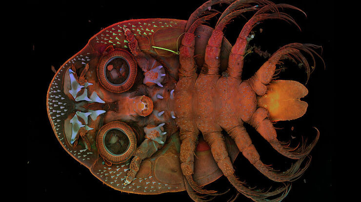
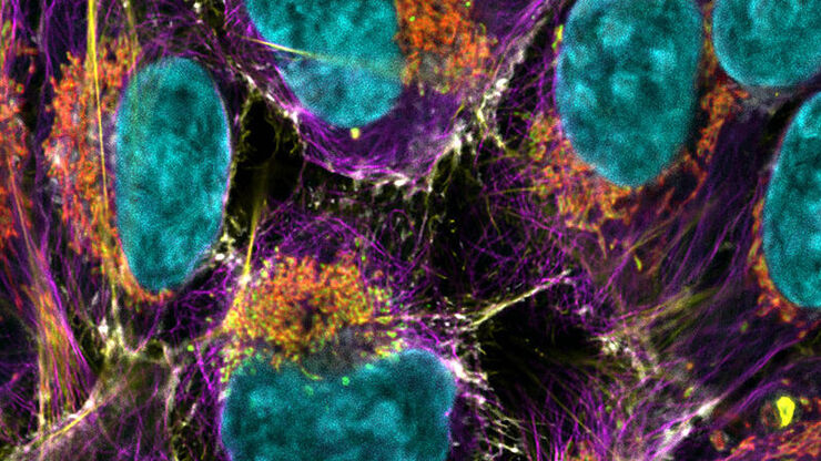
より多くの情報を取得するPotential
STELLARIS独自の TauSense テクノロジー は、全てのサンプルからより多くの情報を抽出し、研究の科学的インパクトを向上させることができます。TauSenseは、蛍光寿命に基づくアプリケーション指向のイメージングツールで構成されており、細胞内の分子の機能を探求するために使用できます。
- TauContrastは、蛍光寿命の変化を使用することで、代謝情報、pH、イオン濃度などの機能情報に対する即座のアクセスを提供します。
- TauGatingは、不要な蛍光の寄与を除去することで画像の品質を向上させます。
- TauSeparationは、スペクトル情報を超えて、実験のおける蛍光シグナルの組み合わせを拡大することができます。
- TauInteractionは、分子間相互作用(たとえば、タンパク質間の相互作用)の検出と定量化を容易にします。
重要なデータへのアクセスを可能にする
Autonomous Microscopy powered by Aiviaで高品質の結果をより迅速に取得することができます。
- STELLARISのライフサイエンス向けのAIベースの希少イベント検出ワークフローは、生物学的サンプル内の希少イベントの最大90%を自律的に検出します。
- 対象物のみを特定して撮影するため、データ取得時間を最大70%短縮します。必要な対象物だけを保存することで、ハードディスク上の貴重なスペースを節約します。
- 顕微鏡観察に費やす時間を節約:自律型の希少イベント検出ワークフローは、通常であればかかる時間を必要としません。
- Autonomous Microscopy powered by Aiviaで、時間の制約や複雑さのために今まで実現が難しかった実験を実行できます。
さらに多くのことを実現するためのProductivity
合理化されたセットアップと画像取得:STELLARISスマートユーザーインターフェイスであるImageCompassは、わずか数回のクリックで複雑な実験も簡単に直感的に設定できます。
- シンプルな早さ:LIGHTNING超解像、ダイナミックシグナルエンハンスメント(DSE)powered by Aiviaにより、リアルタイムで最高の画質を実現します。
- 直感的なユーザーインターフェース: ImageCompassは実験のセットアップから画像取得までをガイドします。
- 実験を最適化する方法:LAS X Navigatorなどのツールはシームレスに統合され、シンプルなイメージングを実現します。
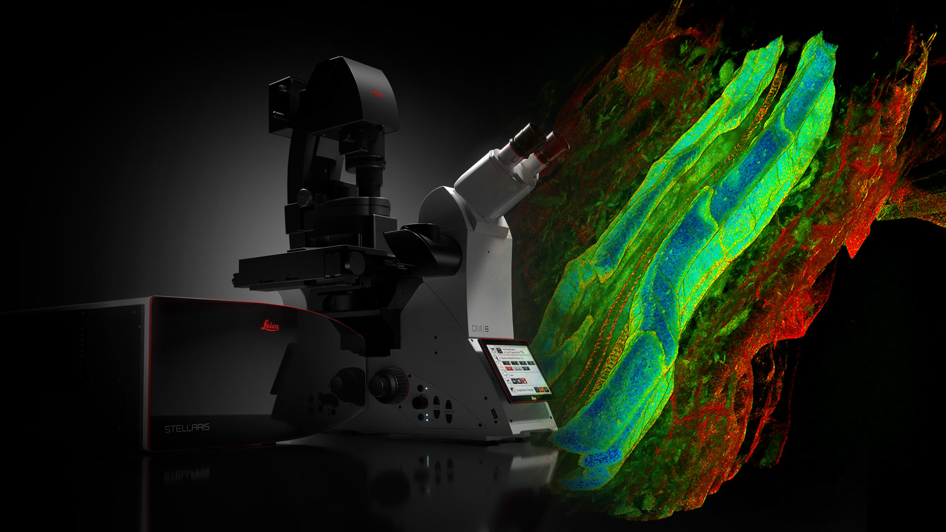
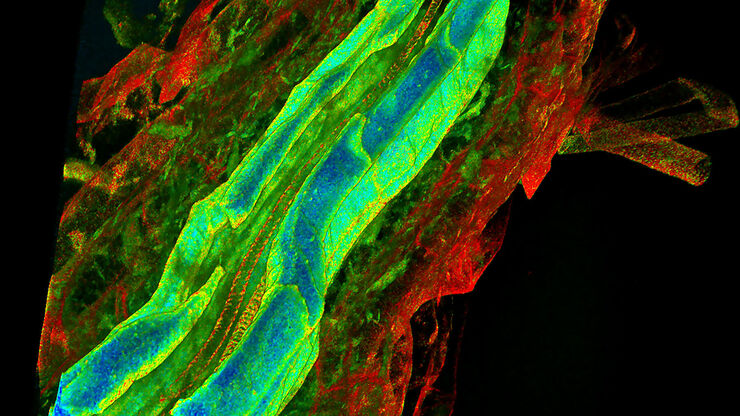
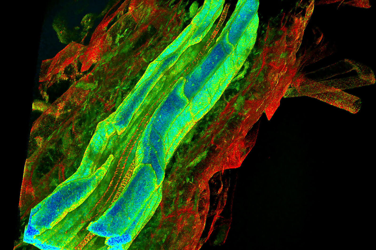
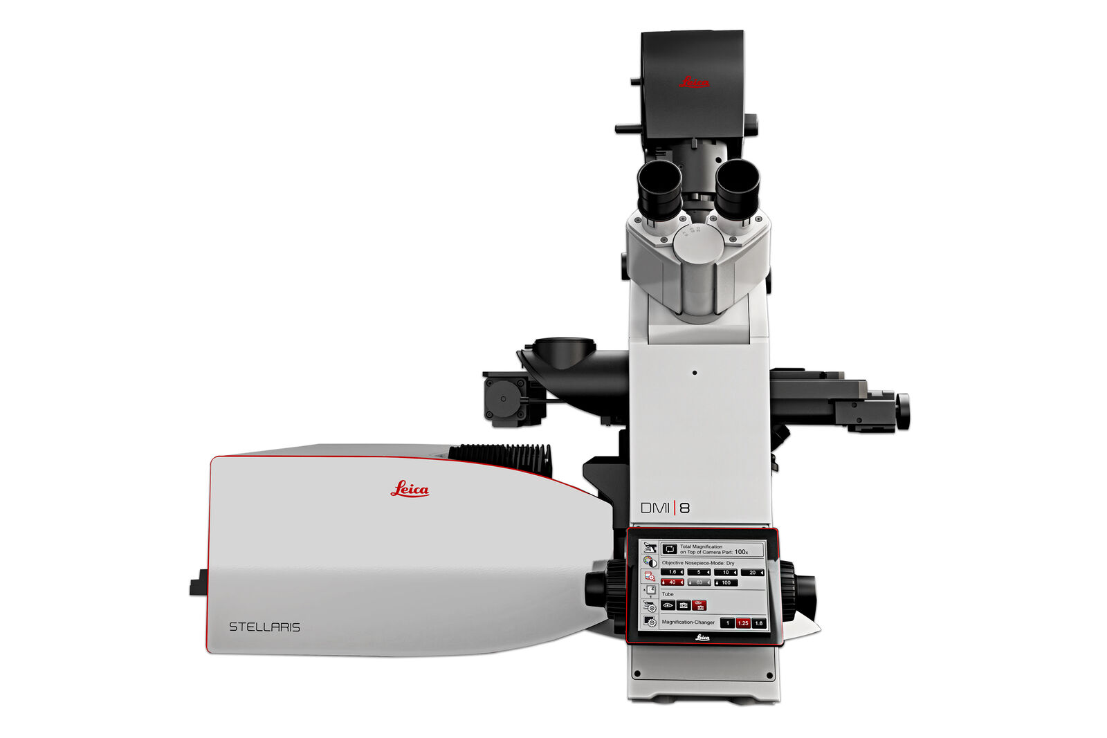
WLLを搭載したSTELLARIS
STELLARISは、ライカの知的財産である音響光学ビームスプリッター(AOBS)とPower HyDファミリー検出器を組み合わせ、さらにWLLを搭載した唯一の共焦点システムです。STELLARISは、独自のTauSenseテクノロジーを統合し、生み出される画像のクオリティと情報量に対する新基準を打ち立てます。
ホワイトライトレーザーを搭載したSTELLARISは、FAst Lifetime CONtrast (FALCON, 蛍光寿命測定)、STED、Deep In Vivo Explorer(DIVE, マルチフォトン)、デジタルライトシート(DLS)、クライオユニット、また、CARS(ラマンイメージング) と組み合わせることができます。STELLARISの新機能は、これらのモダリティーの可能性を最大化し、研究のための新基準を打ち立てるPowerとPotentialを提供します。
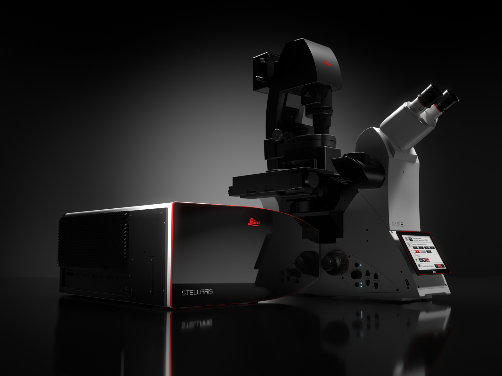
STELLARIS
STELLARISは、スペクトル検出、フォトンカウント検出器、共焦点超解像を提供します。
マルチチャンネル共焦点イメージングは、実験のセットアップと取得を直感的にガイドするスマートユーザーインターフェイス ImageCompass を使用して簡単に達成できます。
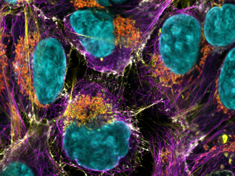
より詳細を観察するためのPower
Power HyD検出器 ファミリーは、より高い光子検出効率 (PDE) *、極めて低いダークノイズ、410 から850 nmの高感度スペクトル検出を提供します。
- 画質を向上:明るさ、解像度、コントラストの理想的な組み合わせ。
- 完全なスペクトルの自由度:次世代ホワイトライトレーザーでは、最大8本の単一励起光をスペクトル全体にわたり同時に使用できます。あらゆる共焦点プラットフォームよりも多くの蛍光色素の組み合わせを用いてイメージングすることができます。NIR領域まで拡張して、より多くの蛍光標識を同時に使用することができます。
- 細胞障害を抑えた穏やかな生細胞イメージング:必要最小限の励起光量で効率的にシグナルを取得することで、サンプルの状態を維持することができます。
* 従来のマルチアルカリ光電子増倍管(PMT)に比べて光検出効率(PDE)を 2 倍に向上、拡張赤色域において 3 倍に向上。