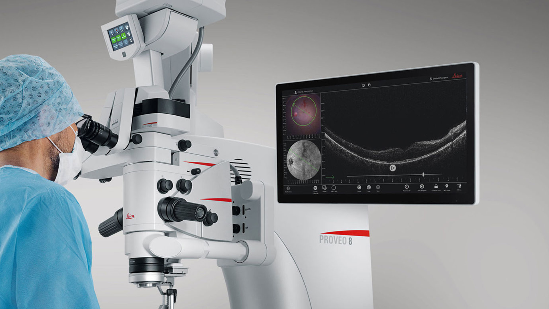
EnFocus 術中 OCT イメージングシステム
ライカ 顕微鏡のOCTソリューションは、眼科手術時において 術者の技能を存分に引き出すためのサポートを提供します。
EnFocus 術中光コヒーレンス トモグラフィ(OCT)により、表層下の組織を観察することが可能です。表層下の組織が外科的手技へどのように反応するか、必要な追加情報をリアルタイムで得ることができます。
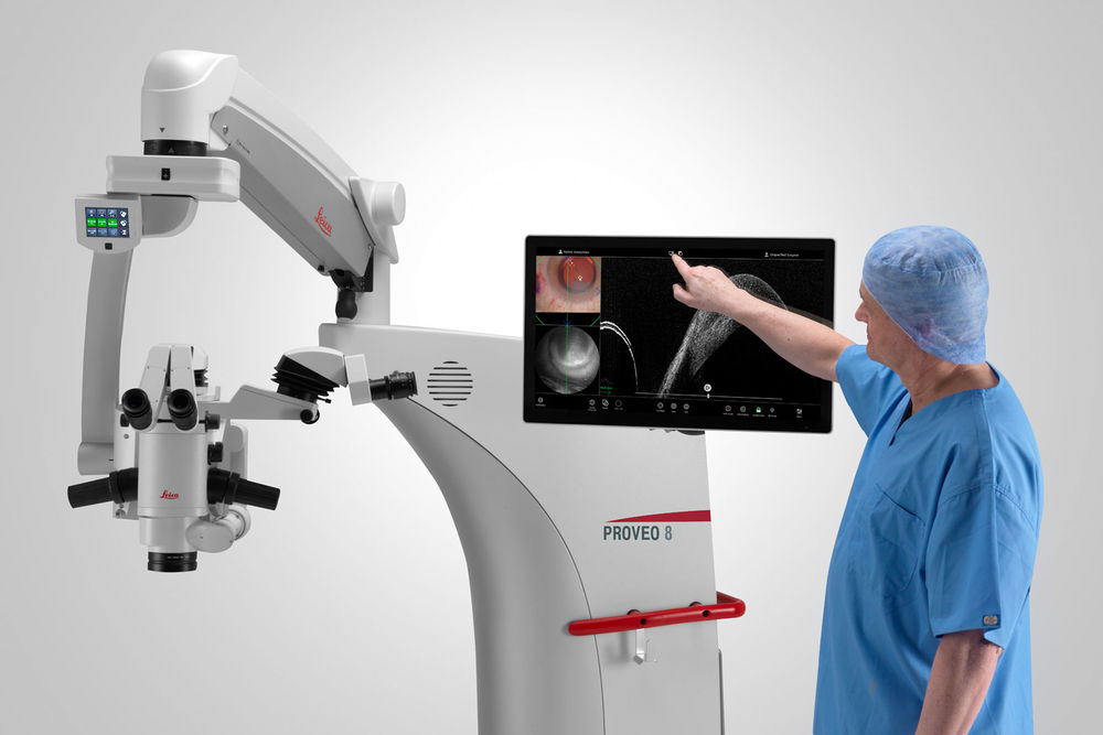
この術中 OCT イメージングシステムは、Proveo 8 眼科手術用顕微鏡の本体内に格納されています。手術中のどの段階においても、数回タップするだけで顕微鏡観察像に OCT イメージングを追加して観察することができます。EnFocus OCT 搭載型 Proveo 8 顕微鏡により、術者は患者様の処置に集中することができます。以下の特徴によりサポートします。
製品またはサービスの承認または提供状況は市場によって異なります。また、認可ラベルおよび指示が国によって異なることがあります。詳細については、お取引ディーラーまでお問い合わせください。

手術のメリット
網膜手術のメリット
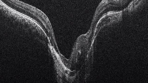
OCTを使用して、膜剥離の張力レベルをリアルタイムで評価し、潜在的な裂傷を回避し、下部にある組織を守ります。 4 µm以下の高分解能画像は、網膜形態の残存膜や黄斑円孔、網膜下浮腫などの合併症の検査にも有用です。
後眼部手術のその他の利点には、次のものがあります:
- フットスイッチを介したスキャンコントロールにより、膜組織に合わせてスキャン角度を迅速に調整します。
- 同期フォーカスを備えたOCULUSのBIOM5などの眼底観察システムに簡単に統合できます
角膜手術のメリット
DMEK(Descemet's Membrane Endothelial Keratoplasty)やDSEAK(Descemet’s Stripping Endothelial Automated Keratoplasty)などの高度な角膜手術では、術者がドナー組織の正しい方向を確認するのをサポートし、再手術を回避するのに役立ちます。
緑内障手術のメリット
最大幅20mmのOCTスキャンは、XENゲルステントの正確な配置をサポートするためのビューを提供します。
OCTは、シャントの配置や、眼圧をコントロールするためにチューブをどの程度固定すべきかのアセスメントに有用です。

EnFocus では、角膜全体と前房をカバーするワイドスキャンが可能です。細部がよく見え、前眼部の細部を可視化します。EnFocus では三次元、すなわち垂直軸 Z 方向での観察が可能になります
即座に確認: 組織の変化と反応を観察
術中に外科の手技に眼組織がどのように反応するのか、リアルタイムに確認することが可能です。必要に応じて手術プランを変更し、手術を進めることができます。
組織の変化と反応を即座にリアルタイムで観察
- 30 fpsのリアルタイム表示により、例えば角膜内皮移植(DMEK/DSAEK)手術においてドナーの組織の密着度を確かめる場合など、各段階で即座のフィードバックが得られます。
- 顕微鏡観察像では確認ができなかった出血などによる合併症が、OCT によって認められた場合、即座に手術プランを変更することができます。
- 再確認のために、取得済みスキャン画像をコマ送り、または再生ビデオモードで簡単に再生し、レビューすることができます。
- たとえば深層層状角膜移植(DALK)手術時の角膜の厚さやニードルの深さなどは、画面上のライブ測定によって再確認できます。

より詳細に観察:表層下の細部をさらに 詳細に観察
これまで隠れていた表層下の組織の細部を明るく、鮮明にイメージングすることで、顕微鏡観察像を補完します。術中 OCT イメージングを一体化し、手術中に眼の病態をより詳細に把握するのに必要な追加的な情報が得られます。
-
- 分散補償ソフトウェアを使用した分光器テクノロジーと、より多くのシグナルを捕捉する高感度の検出器により、アーティファクトと組織を明確に判別できます。
- 組織内の 2.4 μm の軸(深度)方向解像度により、外傷による出血がある場合でも微小な細部を観察できます。
- 最大 1000 A-scan x 1000 B-scan の高いスキャン密度により、高い横方向解像度で網羅的なエリアのスキャンを取得でき、重要な細部を見逃しません。
- 取得した OCT スキャンをコマ送りまたはビデオモードで再生することで、網膜下液が残存しているか、緑内障排液装置 (glaucoma drainage device: GDD)が正しい位置にあるか、角膜移植片はレシピエントの角膜にうまく対置されているかなどの確認が行えます。

表示。顕微鏡画像(左上)、正面像(左下)、OCT B-scan 画像(右)。
より詳細に観察:さらにワイドな観察像
術野全体を確実に観察でき、周辺部の様子を見逃しません。EnFocus により、高倍率でも 20 x 20 mm の横方向視野が得られます。
手術中に観察視野を調整する必要がありません。そのため、眼の中央部で作業している場合でも、周辺部で変化が生じていないかモニターすることができます。

より詳細に観察:臨床的評価から 術中の判断まで
EnFocus OCT を使用することで、術前評価と、術中の組織微細構造の変化に対するリアルタイムの評価で生じるギャップを埋めることができます。
臨床の場では、術中OCT は診療の標準となりつつあります。OCT イメージングシステムは、手術室内での使用がますます増えており、術中の判断に大きく関わっています。DISCOVER 調査* では、術中 OCT の利用による影響に関するデータが収集されました。この調査で明らかになったのは、顕微鏡と OCT の併用により追加情報が得られ、それに基づいて術中に手術プランを変更できることです。また、膜剥離手術において 35% の事例で、術者の当初の印象と術中 OCT による知見とが違ったことが明らかにされています。
*View data from the DISCOVER Study: 顕微鏡に統合された術中 OCT についての調査(DISCOVER)
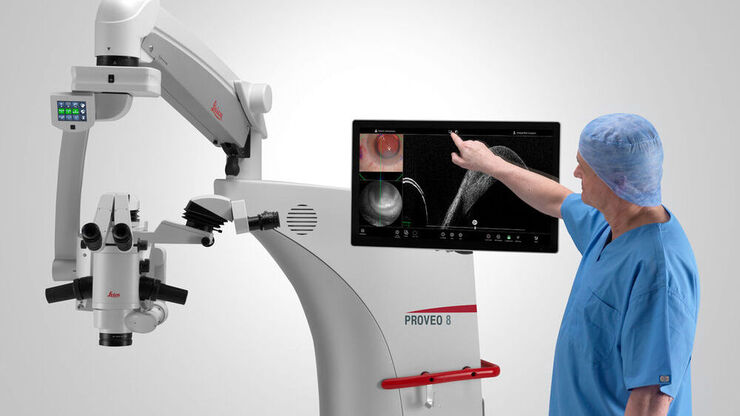
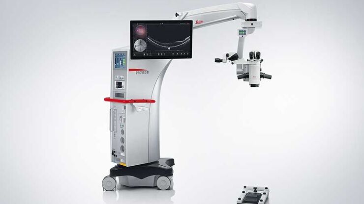
最大限の自由度:スムーズで独立した ワークフロー
EnFocus を Proveo 8 に組み込むことで、ワークフローがサポートされ、OCT 画像を独立して操作できるようになります。術者は手術に専念することができます。.
- フットスイッチ、ハンドル、または 27 インチタッチスクリーン HD モニターを使って、手術中のどの段階でも自分で簡単に術中 OCT を有効にすることができます。
- 手術前に術式とステップに応じて個人設定とモードをあらかじめプログラムしておき、フットスイッチまたはハンドルに割り当てることで、スムーズなワークフローを実現します。
- 同様にして画像を記録・取得し、Med X Change の Evolution4K 録画システムを選ぶことで、網羅的な記録が可能です。
- DI C800 を使って顕微鏡画像と術中 OCT 画像を接眼レンズ内に視野内表示したり、27 インチ HD タッチスクリーンモニター上に表示したり、4 つのビデオ出力を利用して大型スクリーンに投影することも可能です。
手術室内におけるワークフローの自由度
網膜硝子体手術に携わる Dr. Barbara Parolini(イタリアの Eyecare Clinic Brescia)が、EnFocus OCT イメージングシステムが手術室内にどのような自由をもたらすか語ります。
