お問い合わせ

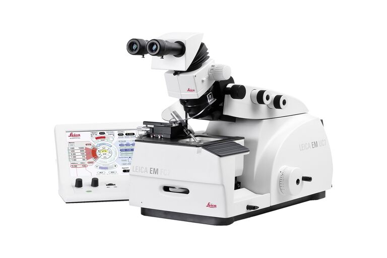
Leica EM UC7 常温・凍結切片作製用 ウルトラミクロトーム

最新の記事を読む

Water Flea Daphnia imaged by Electron Microscopy. Courtesy of Mag. Dr. Gruber, University of Vienna, Austria

Streamline your EM Sample Preparation Workflow for Biological Applications

Master EM sample preparation, including ultramicrotomy, for life sciences in this expert eBook!

Studying Virus Replication with Fluorescence Microscopy

The results from research on SARS-CoV-2 virus replication kinetics, adaption capabilities, and cytopathology in Vero E6 cells, done with the help of fluorescence microscopy, are described in this…
Acrylonitrile butadiene styrene (ABS) stained with osmium tetroxide (OsO4), sectioned with a DIATOME diamond knife at room temperature, and then imaged with HAADF TEM.

Ultramicrotomy Techniques for Materials Sectioning

Learn about ultramicrotomy for materials sectioning when investigating polymers and brittle materials with transmission (TEM) or scanning electron microscopy (SEM) or atomic force microscopy.
Micrograph of dinoflagellate cells. Scale bar = 1 µm.

How Marine Microorganism Analysis can be Improved with High-pressure Freezing

In this application example we showcase the use of EM-Sample preparation with high pressure freezing, freeze substiturion and ultramicrotomy for marine biology focusing on ultrastructural analysis of…

Exploring the Structure and Life Cycle of Viruses

The SARS-CoV-2 outbreak started in late December 2019 and has since reached a global pandemic, leading to a worldwide battle against COVID-19. The ever-evolving electron microscopy methods offer a…

Investigating Synapses in Brain Slices with Enhanced Functional Electron Microscopy

A fundamental question of neuroscience is: what is the relationship between structural and functional properties of synapses? Over the last few decades, electrophysiology has shed light on synaptic…

Workflows and Instrumentation for Cryo-electron Microscopy

Cryo-electron microscopy is an increasingly popular modality to study the structures of macromolecular complexes and has enabled numerous new insights in cell biology. In recent years, cryo-electron…

Microscopy in Virology

The coronavirus SARS-CoV-2, causing the Covid-19 disease effects our world in all aspects. Research to find immunization and treatment methods, in other words to fight this virus, gained highest…

Workflow Solutions for Sample Preparation Methods for Material Science

This brochure presents and explains appropriate workflow solutions for the most frequently required sample preparation methods for material science samples.

Ultrastructural Preservation and Improved Visualization of Membranes in Primary Bovine Chromaffin Cells

Intracellular dynamic events, such as protein trafficking, recycling, and degradation as well as signal transduction, to name a few, can be studied using live-cell imaging techniques. However,…
Ultramicrotomy; ARTOS 3D

Introduction to Ultramicrotomy

When studying samples, to visualize their fine structure with nanometer scale resolution, most often electron microscopy is used. There are 2 types: scanning electron microscopy (SEM) which images the…

Bridging Structure and Dynamics at the Nanoscale through Optogenetics and Electrical Stimulation

Nanoscale ultrastructural information is typically obtained by means of static imaging of a fixed and processed specimen. However, this is only a snapshot of one moment within a dynamic system in…

Superior Ultrastructural Preservation and Structural Contrast in Drosophila Tissue

Optimal structural preservation of tissue can only be obtained by high pressure freezing. However, preparing the samples in optimal conditions is challenging. This article explains in detail how to…

Cryo-Electron Microscopy of Vitreous Sections (CEMOVIS) of Yeast

Application Note for Leica EM HPM100 - The sections are of yeast frozen with a Leica HPM100 high pressure freezer in the copper tube system, the cell paste was mixed with a pH 6.5 MES/dextran buffer…

Perusing Alternatives for Automated Staining of TEM Thin Sections

Contrast in transmission electron microscopy (TEM) is mainly produced by electron scattering at the specimen: Structures that strongly scatter electrons are referred to as electron dense and appear as…

Atomic Force Microscopy Study of a Stretched Impact Copolymer

Atomic force microscopy (AFM) is a powerful tool in the suite of nanoscale characterization techniques that provides a variety of information including topography, mechanical properties, and…

応用分野

ゼブラフィッシュを用いた研究

スクリーニング、ソーティング、マニピュレーションおよびイメージングを通じて最良の結果を得るためには、細部や構造を観察して、研究の次の段階に向けて正しい判断を下す必要があります。 優れた光学系と高解像度で定評のあるライカの実体顕微鏡と透過照明スタンドは、世界中の研究者から支持されています。
Background image
Scroll to top