Leica 
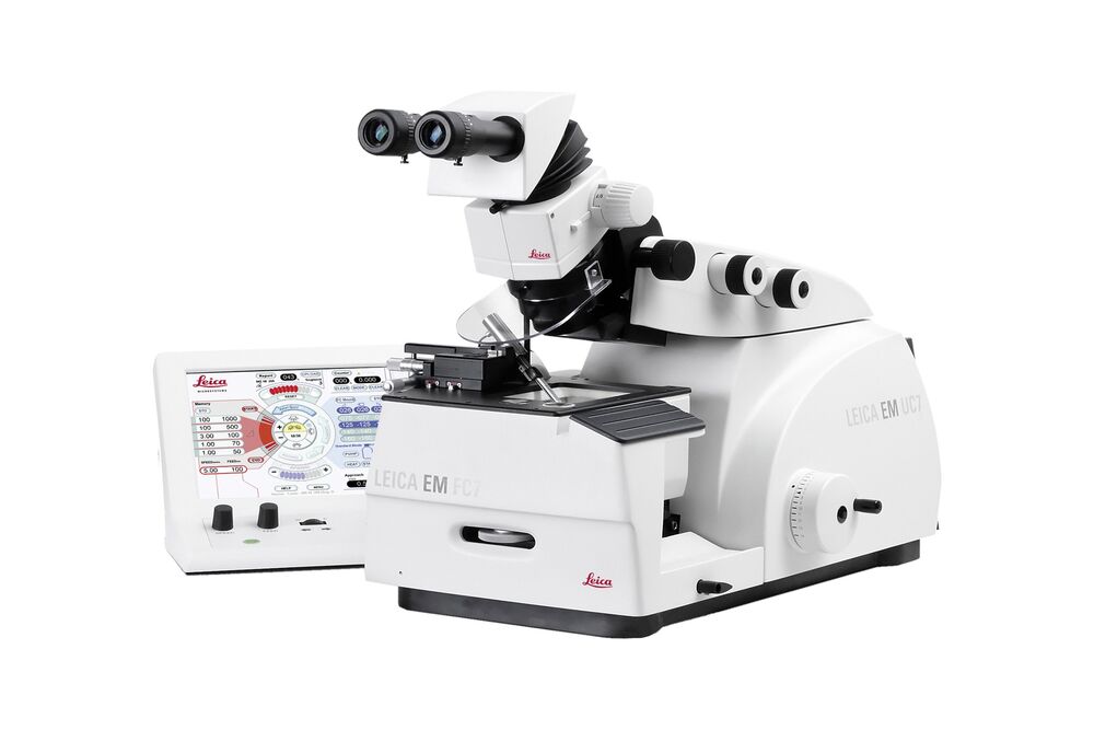
EM UC7 常温・凍結切片作製用 ウルトラミクロトーム<br>
光学顕微鏡(LM)、電子顕微鏡(EM)、および原子間力顕微鏡(AFM)の観察で用いる高品質切片や試料断面を、これまでになく簡単にかつ正確に作製できる先鋭の試料作製技術を搭載。
ライカEM UC7は、LMで用いる準超薄切片から、TEMで要求される高品質な超薄切片、さらにSEM、あるいはAFM 観察で必要な高い面精度の断面作製にも活用できます。超精密機構でありながら、人間工学に基づいた設計、およびタッチパネル式コントロールユニットの直感的レイアウトを採用したライカEM UC7は、様々な場面で最高品質の試料作製に最適化されています。
Benefits
- Obtain applicable, reproducible results by preparing high-quality consistent ultrathin sections with a thickness feed range from 1 nm to 15 µm
- Benefit from an optimal knife approach to the sample, even at low water levels, with our patented eucentric movement
- Avoid unnecessary loss of sample material thanks to the optimal positioning of the optical head with outstanding LED illumination giving superior visibility of the knife edge and block face
- Save valuable time with the fully motorized knife stage featuring North-South and East-West movement using the autotrim mode and measuring function.
Leica Science Lab: Superior Ultrastructural Preservation and Structural Contrast in Drosophila Tissue
Prepare consistent, thin sections with ease
Access all sectioning parameters easily with the ergonomic touchscreen control unit of the EM UC7 ultramicrotome. Quickly adjust feed, cutting speed, and illumination parameters during the sectioning process on the main screen of the unit. The control wheels help you to set the East-West and North-South movement of the knife stage accurately, making the knife approach to the sample block easy and fast. Prepare perfect block faces with the auto-trim function, thus, ensuring consistent parallel edges of the attained sections.
- Full control over all sectioning parameters with the ergonomic touch-screen control panel
- User defined quick access presets
- High reproducible sectioning capabilities thanks to accurate feed settings of the fully motorized knife stage and auto-trim function
- Cutting speed range from 0.05 to 100 mm/s
- Vibration-decoupled gravity-stroke cutting arm for chatter-free sectioning
Save valuable sample material
With the patented eucentric movement of the EM UC7’s microscope carrier, you can rest assured to save valuable sample material. The function provides the proper illumination and a perfect viewing angle of the glass- and diamond-knife edge. It allows the examination of sections, even with lower water levels. The defined eucentric microscope carrier position increases the approach accuracy whilst ensuring reduced sample material loss, particularly crucial during mono-layer sectioning.
Prepare your samples with superior visibility
Make approach and alignment to the sample block fast, easily, and safe! The EM UC7 ultramicrotome is equipped with three independent, built-in, brightness-controlled LED light sources and additional LED spotlights. Benefit from outstanding illumination of the sample block and knife edge during the whole preparation process.
- Optimal LED illumination for top, back, and transmitted light
- Superior visibility through a focused light beam of the LED spot illumination
- All illumination modes can be controlled independently
Save time with automatic trimming
Free up time in the laboratory with the automatic trimming function of the EM UC7 ultramicrotome. Via the specialized function of the touch-screen control panel, the block face can be trimmed with minimal user interaction. The fully motorized knife stage ensures an extremely precise movement of the knife to the sample in a North-South and East-West direction. Confidently operate the instrument without the risk of damaging the sample block or the knife!
- Save valuable time with the autotrim function
- Document knife-usage information for perfect thin sections
- Fully motorized and highly accurate East-West and North-South knife-stage approach for reproducible results
- Measure the size of the sample block face
Expand your EM UC7 to a cryo sectioning system
Equip your EM UC7 Ultramicrotome with the EM FC7 cryochamber and make it a state-of-the-art cryo sectioning system in just a few minutes. Samples mounted in the EM FC7 allow for preparation of ultrathin cryo-sections at temperatures between -15°C and -185°C. Easily section a variety of specimens ranging from frozen biological material to polymers and rubbers with the utmost precision in only a few simple steps.
- Three different cryo-modes: 1) standard, 2) high gas flow, and 3) wet cryo-sectioning
- Highly stable temperature regulation and low LN2 consumption
- Electrostatic charge and discharge functions for section adherence with the EM CRION ionizer (optional)
- Attachable micromanipulator for precise positioning of TEM grids for easy section collection (optional)
- Direct transfer of sections to TEM or SEM in cryo-conditions via the EM VCT500 (optional)
Choose your workflow according to your biological question
The EM UC7 ultramicrotome will help you get high quality results when preparing samples for TEM and SEM room temperature workflows. There is also the option for an expansion with the EM FC7 cryo-chamber allowing for Tokuyasu and CEMOVIS applications. For more information on workflow solutions for your research, download our workflow booklets
Post-fixation epoxy CLEM
This workflow identifies the structure of interest in tissue samples by using a mild chemical fixation, followed by LM imaging. Afterwards, the sample is further processed with electron microscopy fixatives, then embedded, sectioned, stained, and analyzed in a TEM.
Hybrid Processing
This standard workflow, which is done before TEM analysis, combines: 1) superior ultrastructural fixation with high-pressure freezing and freeze-substitution embedding, 2) room temperature manipulation via trimming, 3) ultrathin sectioning, and 4) staining.
ライカまでお気軽にご相談ください