お気軽にお問い合わせください
細胞解析アプリケーション向けソリューションの専門家が、最適なアドバイスでサポートいたします。

細胞メカニズムは病気の進行にどのような影響を及ぼすのか?
細胞レベルでの仕組みを理解することは、がん研究やアルツハイマー病などの分野において極めて重要です。例えば、細胞シグナル伝達経路はがん細胞の増殖を制御し、一方で特有の細胞変化がアルツハイマー病の病態形成に関与しています。

ライブセル解析に最適な手法とは?
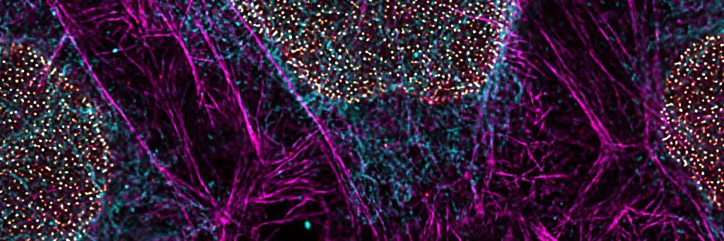
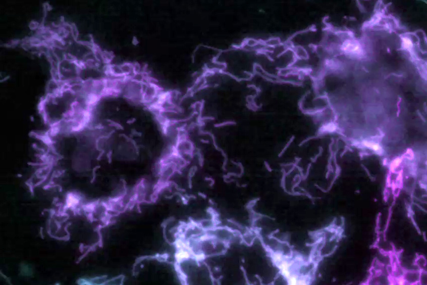
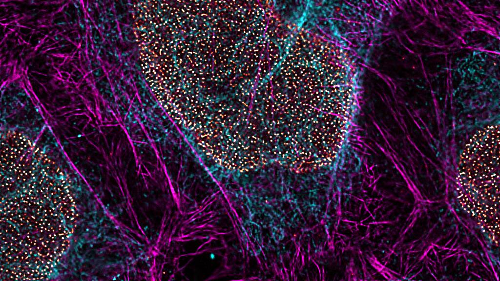
ワイドフィールド、共焦点、多光子、さらには超解像顕微鏡といった先進的なイメージング技術により、生細胞のダイナミクスをリアルタイムで可視化することが可能になります。これらの技術により、細胞のダイナミクスや挙動を高精度に捉えることができ、幅広い細胞解析研究を強力に支援します。

どのようにして包括的な細胞解析を実現できるのでしょうか?
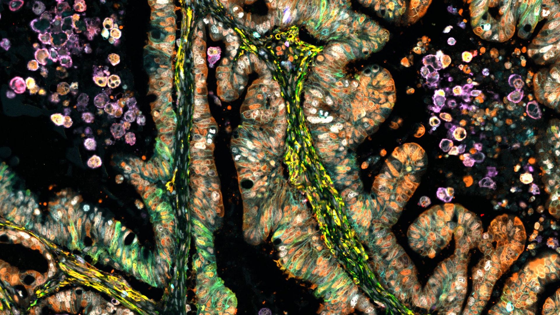
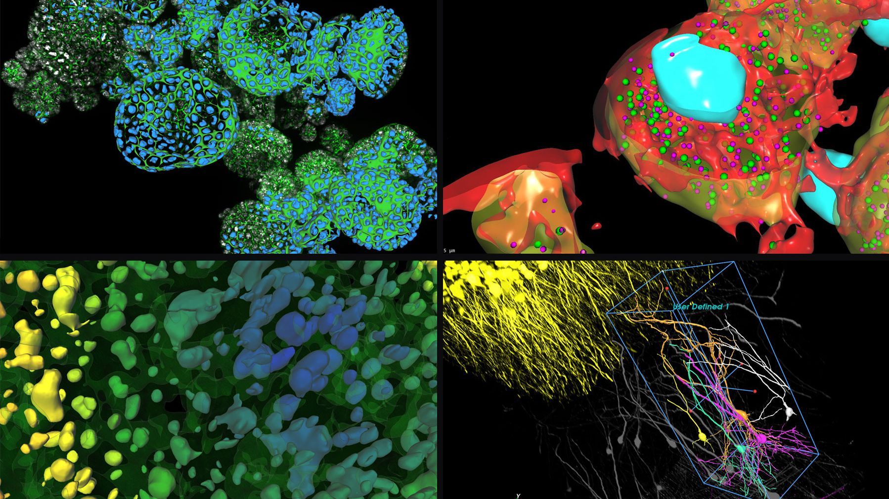
機能イメージングや相関イメージングなど、多様なイメージング手法を統合することで、より高度で包括的な解析が可能になります。このアプローチにより、細胞内プロセスや相互作用への理解が一層深まります。
Leicaのソリューションは、細胞解析に取り組む研究者をどのように支援するのでしょうか?
関連記事

Researchers Insights: Microscopy in Cancer Research
Discover how imaging techniques are driving cancer research forward. In this issue, we present comprehensive multimodal studies using microscopy, as well as new directions in intraoperative cancer…
 and astrocytes (green) in a cortical spheroid derived from human induced pluripotent stem cells.")
Guide to Live-Cell Imaging
For a wide range of applications in various research fields of life science, live-cell imaging is an indispensable tool for visualizing cells in a state as close to in vivo, i.e. living and active, as…

Precision and Efficiency with AI-Enhanced Cell Counting
This article describes the use of artificial intelligence (AI) for precise and efficient cell counting. Accurate cell counting is important for research with 2D cell cultures, e.g., cellular dynamics,…

Live-Cell Imaging Techniques
The understanding of complex and/or fast cellular dynamics is an important step for exploring biological processes. Therefore, today’s life science research is increasingly focused on dynamic…
 inoculated with cowpea mosaic virus (CPMV) containing the GFP-gene inserted between the movement protein (MP) and the capsid proteins (CPs) in the viral RNA 2")
Introduction to Live-Cell Imaging
The understanding of complex and fast cellular dynamics is an important step to get insight into biological processes. Therefore, today’s life science research more and more demands studying…
