がん免疫学における3Dハイプレックスイメージング。マウスモデルの膵腫瘍切片の詳細。15種のマーカーで標識し、STELLARIS SpectraPlex によりワンショットで撮像。総撮像体積:3.2 mm × 2.2 mm × 70 µm、撮像時間:1時間20分。出典:3D high-multiplex imaging in cancer immunology.Kunz L., Speziale D., et al., Nat.Methods(2024年)。

高度な組織イメージングおよび解析

生物学的過程および疾患機構を解明するために、研究者は細胞と細胞外成分の相互作用を理解すべく、組織を多層的に解析します。先端的なイメージングおよび解析ソリューションは、組織の高精細な可視化と包括的解析を提供し、空間生物学研究を加速させます。

お気軽にご連絡ください!

高度な組織イメージングおよび解析のソリューションに関するアドバイスを、当社のイメージングエキスパートがご提供します。

TauSTED Xtend により取得した画像。3色のSTEDサンプル。シアン:ビメンチン、AF594マゼンタ:ファロイジン-アクチン、Atto 647N。グロー効果:NUP 107, CF680R.サンプル提供:Dr. Mariano Gonzales Pisfil and Dr. Steffen Dietzel,  Core Facility Bioimaging, Biomedical Center,  Ludwig Maximilian University of Munich, Germany

あらゆるアプリケーションに応える汎用性

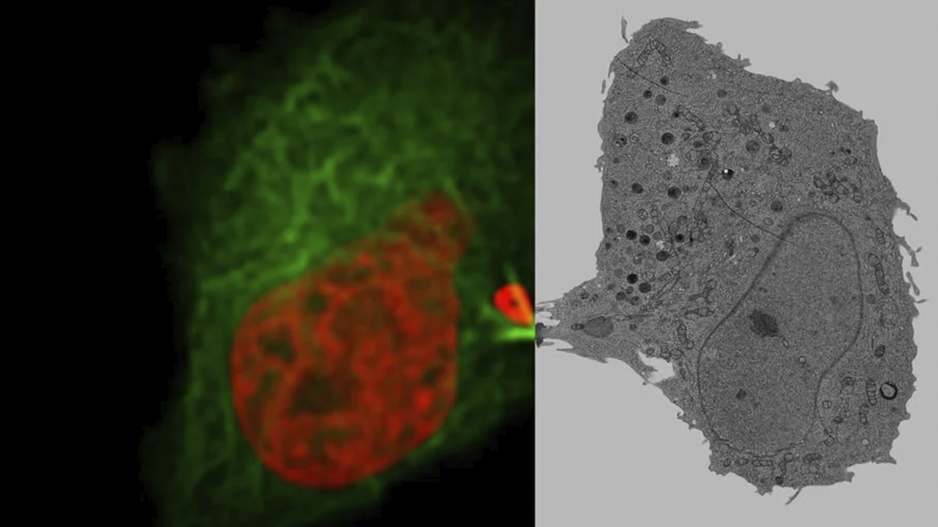
2D/3D の組織データ取得に対応するため、ワイドフィールドによる広範囲観察から、超解像共焦点顕微鏡による微細構造解析まで、多様なイメージングソリューションを提供しています。この汎用性によって、組織構造および機能を効率的かつ的確に解析するための適切なツールが研究者に提供されます。

先進的マルチプレックス解析とフェノタイピング

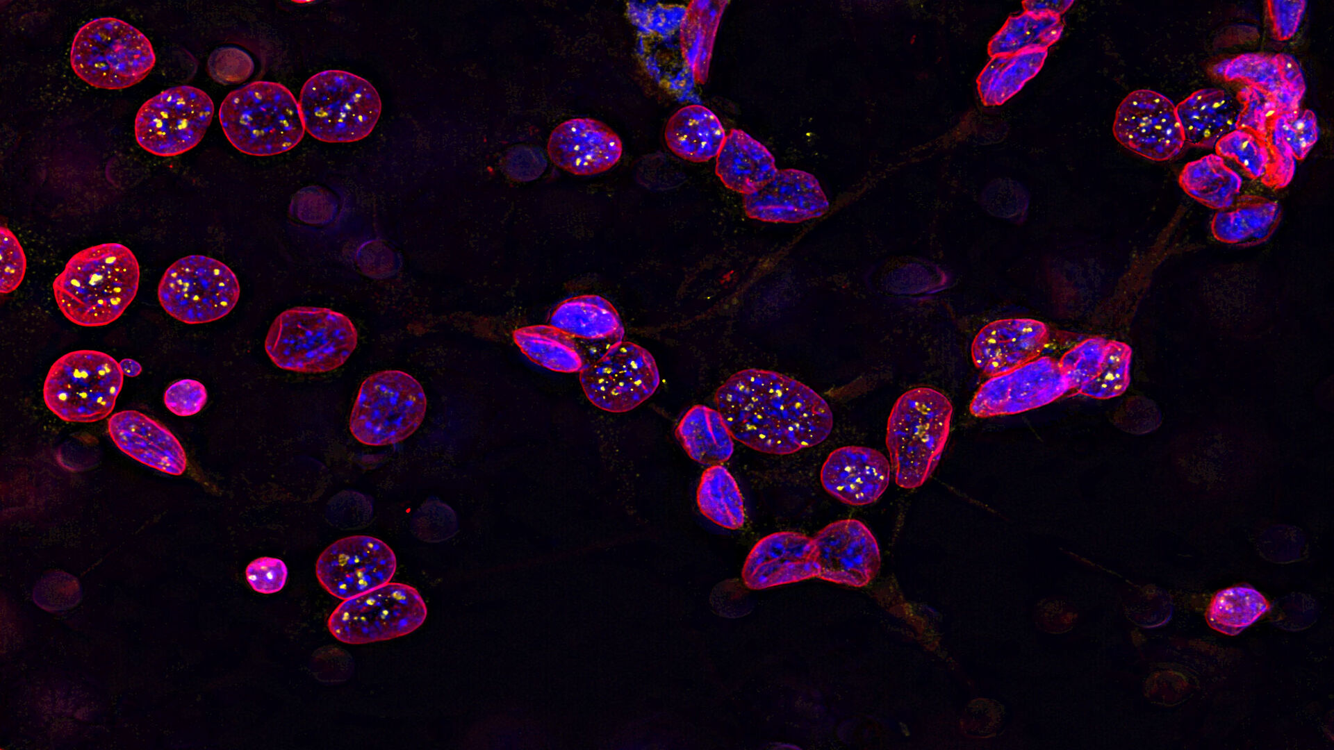
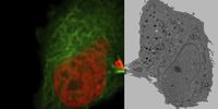
空間生物学のマルチプレックス解析およびフェノタイピングにおいて、当社のマルチプレックスソリューションを活用することで、組織サンプル中の複数バイオマーカーを包括的に解析できます。これらの手法には、単一サンプルにおいて60種類以上のバイオマーカーを解析できる自動反復染色が含まれており、複雑な生物学的プロセスや相互作用の解明において重要な役割を果たします。

包括的な組織スキャンソリューション

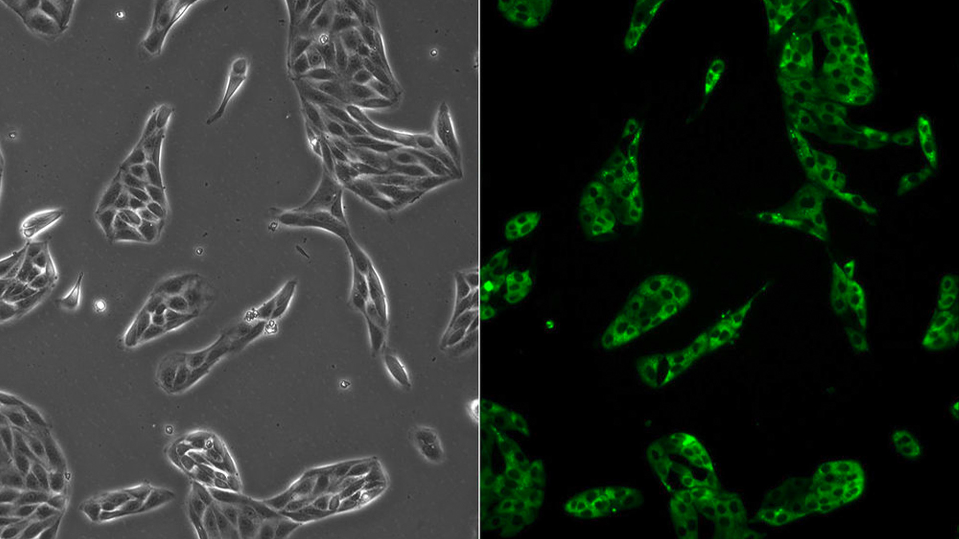
薄切片およびボリュームスキャンに対応した多機能機器を含む当社のソリューションは、さまざまな組織解析アプリケーションを支援します。この柔軟性によって、組織構造および機能の解析に最適なソリューションを効果的に適用することが可能となります。

組織イメージングおよび解析に Leica 顕微鏡ソリューションを用いる利点とは何でしょうか?


組織の構造および機能を研究する意義は何でしょうか?

科学者は、組織、細胞、分子間の関係性を解明するため、異なるスケールにわたって組織構造および機能を解析します。Leica のイメージングソリューションとサンプル前処理技術は、細胞をフェノタイプ別に識別することを可能にし、その知見を健康および疾患への理解に応用することができます。


組織機能の解析に最も適した手法は何でしょうか?

組織機能や多様な刺激に対する応答を理解するためには、組織内の生物学的プロセスを可視化・解析することが不可欠です。Leica の機能イメージングおよび空間フェノタイピングツールは、こうした目的の達成を効果的にサポートします。


再現性のある結果を迅速に取得するためには、どのような手法が有効ですか?

標準化されたワークフローと自動化イメージングに、AI ベースの解析ソフトウェア Aivia を組み合わせることで、データ処理の高速化、エラー低減、一貫した高品質な解析結果が実現し、信頼性と再現性の高い成果が得られます。

高度な組織イメージングと分析に関するよくある質問

Show answer 創薬や医薬品開発において、なぜ生死判別アッセイを活用するのでしょうか?

患者の安全が最優先です。従って、致死量に関する知識は非常に重要であり、臨床試験を検討する前に規制当局から要求されるのです。生死判別アッセイを用いることで、閾値を評価することが可能となります。十分な量のデータを効率的に取得するために、Micaは最適なソリューションです。

Show answer レーザーマイクロダイセクションによって、バイオ医薬品研究はどのように改善されるのでしょうか?

高精度に組織領域を回収することで、高品質のゲノムおよびプロテオーム解析を可能にします。レーザーマイクロダイセクションは、バイオ医薬品研究者が特定の細胞や組織領域をターゲットとし、医薬品開発においてより正確でコンタミネーションのない研究を促進するのに役立ちます。

Show answer 創薬や医薬品開発に関するバイオファーマの研究になぜ顕微鏡を使うのでしょうか?

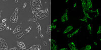
顕微鏡観察は、化合物や薬剤候補に対する細胞応答の詳細な空間的知見を得るのに役立ちます。創薬段階、すなわち生細胞培養モデルや組織の品質管理や反応経路の詳細な分析には、さまざまな創薬・開発ニーズに対応するライカ製品があります。

関連記事

高度な組織イメージングおよび解析に関する最新記事を読む

ライカ マイクロシステムズの知識ポータルでは、顕微鏡に関する科学的な研究記事および教材を提供しています。日常業務や研究で、ビギナーの方から経験豊富な方まで幅広くサポートします。

その他の記事

History, Developments and Trends of Microscopy in Cancer Research

Cancer is a global disease, with 18 million new cases diagnosed and 10 million cancer-related deaths worldwide in 2020. This burden is set to increase, with a projected increase in cases of ~55% by…

Researchers Insights: Microscopy in Cancer Research

Discover how imaging techniques are driving cancer research forward. In this issue, we present comprehensive multimodal studies using microscopy, as well as new directions in intraoperative cancer…
Final Segmentation of organelles in Trichomonas species. Magenta – costa, light blue – hydrogenosomes, turquoise – ER, red – vacuoles, yellow – axostyle, green – Golgi apparatus.  Sample courtesy of Isabelle Guerin-Bonne, Low Kay En, Electron Microscopy Unit, Yong Loo Lin School of Medicine, National University of Singapore. Scale bar: 1 µm.

Volume EM and AI Image Analysis

The article outlines a detailed workflow for studying biological tissues in three dimensions using volume-scanning electron microscopy (volume-SEM) combined with AI-assisted image analysis. The focus…
Pancreatic Ductal Adenocarcinoma imaged with Cell DIVE. Analysis done by Aivia.

A Guide to Spatial Biology

What is spatial biology, and how can researchers leverage its tools to meet the growing demands of biological questions in the post-omics era? This article provides a brief overview of spatial biology…
Masson-Goldner staining of a hedgehog brain slice.

How to Streamline Your Histology Workflows

Streamline your histology workflows. The unique Fluosync detection method embedded into Mica enables high-res RGB color imaging in one shot.
Spectral separation of 11 fluorophores coupled to polystyrene beads on a STELLARIS confocal system.

Multiplexing through Spectral Separation of 11 Colors

Fluorescence microscopy is a fundamental tool for life science research that has evolved and matured together with the development of multicolor labeling strategies in cells tissues and model…
3D Reconstruction of brain slide image_Mica

3D Tissue Imaging: From Fast Overview To High Resolution With One Click

3D Tissue imaging is a widespread discipline in the life sciences. Researchers use it to reveal detailed information of tissue composition and integrity, to make conclusions from experimental…

Tissue Image Gallery

Visual analysis of animal and human tissues is critical to understand complex diseases such as cancer or neurodegeneration. From basic immunohistochemistry to intravital imaging, confocal microscopy…
14: 7-day old 3D cell culture of MDCK cells stably transfected with; Mx1-GFP (green), SPY555-Actin (yellow) and WGA-AlexaTM 647 (magenta), and imaged using a THUNDER Imager Cell Spinning Disk. Shown are both the spinning disk image (left) and the same image after THUNDER Computational Clearing was applied (right); scale bar 50 μm.

オルガノイドと3D細胞培養

ライフサイエンス研究で近年最も目覚ましい発展の一つは、オルガノイド、スフェロイド、生体機能チップモデルなどの3D細胞培養システムの開発です。 3D細胞培養は、細胞が成長し、全3次元で周囲と相互作用できる人工環境です。 これらの条件は、生体内条件に似ています。

もっと読む

Image of C2C12 cells: The cells are stained with lamin B (magenta) which indicates nuclear structure, Hoechst (blue) indicating DNA, and γH2AX (yellow) indicating damage to DNA. Cells were imaged using a THUNDER Imager 3D Live Cell with a 63X/1.4 oil immersion objective.

細胞生物学研究

細胞生物学イメージングは、さまざまな光電子相関顕微鏡を使用して行われます。 ライカマイクロシステムズが提供するイメージングソリューションは、細胞生物学研究を最大限に行えるように設計されています。

もっと読む

Mammalian cells expressing H2B-mCherry and alphaTubulin-mEGFP to visualize DNA and microtubules respectively. Left: Widefield imaging using THUNDER Imager. Right: Sample after correlated EM imaging using Leica Microsystems Coral Life workflow. Cells were cryo-immobilized by high-pressure freezing 60 min after the onset of cytokinesis. The intercellular bridge was imaged under TEM after freeze substitution and resin infiltration.

光電子相関顕微鏡法(CLEM)

ライカマイクロシステムズ独自のCLEMソリューションは、サンプルの生存性や品質チェック、精密かつ確実な3Dターゲティング機能を確保しています。 光電子相関顕微鏡ソリューションを活用することで、適切な細胞を最良のタイミングで直接識別し、高解像度のクライオ共焦点データを入手し、蛍光情報を微細構造コンテクストで捕らえます。

もっと読む

細胞培養

ラボ環境での細胞培養は、細胞生物学、がん研究、発生生物学をはじめ、ライフサイエンスに関するすべての分野、そして薬学の研究の基本です。ライカが研究室内での細胞培養をどのようにサポートしているか、こちらでご覧いただけます。

もっと読む

14: 7-day old 3D cell culture of MDCK cells stably transfected with; Mx1-GFP (green), SPY555-Actin (yellow) and WGA-AlexaTM 647 (magenta), and imaged using a THUNDER Imager Cell Spinning Disk. Shown are both the spinning disk image (left) and the same image after THUNDER Computational Clearing was applied (right); scale bar 50 μm.
Image of C2C12 cells: The cells are stained with lamin B (magenta) which indicates nuclear structure, Hoechst (blue) indicating DNA, and γH2AX (yellow) indicating damage to DNA. Cells were imaged using a THUNDER Imager 3D Live Cell with a 63X/1.4 oil immersion objective.
Mammalian cells expressing H2B-mCherry and alphaTubulin-mEGFP to visualize DNA and microtubules respectively. Left: Widefield imaging using THUNDER Imager. Right: Sample after correlated EM imaging using Leica Microsystems Coral Life workflow. Cells were cryo-immobilized by high-pressure freezing 60 min after the onset of cytokinesis. The intercellular bridge was imaged under TEM after freeze substitution and resin infiltration.
Scroll to top