
Introduction
Living organisms are intricate and complex. Their complexity is best unraveled by examining the various cells and tissues that govern their anatomy, organs, and physiology. Histology, the science of studying tissues at the sub-cellular, microscopic level, is a cornerstone of biological research. It allows scientists to probe the inner workings of cells, the architecture of organs, and the underlying function of life's building blocks.
The usefulness of histological staining lies in its ability to reveal sub-cellular details of tissues. When a fresh tissue section is prepared in the laboratory without staining, then the images recorded with a conventional microscope using basic illumination lack the necessary contrast needed to reveal critical details. Staining is used to highlight important features of the tissue by enhancing tissue contrast. This improves the investigator's ability to draw conclusions, such as identifying pathological abnormalities.
How is staining done?
Histological staining encompasses a variety of techniques, each with its own set of required compounds and protocols. They can be broadly categorized into two classes: chemical-based and antibody-mediated staining.
Chemical-based staining, as the name suggests, involves at least one step of immersing tissue sections in solutions containing dyes or stains. These chemicals bind to specific sub-cellular structures or substances, thereby imparting color and contrast to the tissue. One of the most common techniques is Hematoxylin and Eosin (H&E) staining which is a staple in histology laboratories worldwide.
In contrast to chemical-based staining, antibody-mediated staining offers a higher flexibility to specifically target structures. Antibodies selectively target specific cellular components or biomarkers of interest. These antibodies or secondary antibodies are often labeled with an enzyme, such as peroxidase, which catalyzes a reaction to produce a visible substrate, thereby creating a localized and distinct signal within the tissue.

How histological samples are imaged with traditional RGB-cameras
Unlike fluorescence, a histologic staining can be seen with the naked eye. In microscopes, it also works with light passing through the tissue, while the contrast comes from the differential blocking of light passing through the specimen. Fluorescence contrast shines light of a specific wavelength, i.e. color, through the same objective onto the specimen and detects the emitted light of a different color. This is why we see bright objects on a dark background, while in histology the contrast is reversed - dark objects on a bright background.
The color information of a pixel is represented by the balance of channel intensities for the red (R), green (G), and blue (B) intensities. However, if we take a closer look at a Bayer matrix sensor, we see that each of the pixels can only detect one of the three colors. This is determined by the filter in front of a given pixel. Therefore, the discrete R, G, and B information must be merged into a combined RGB color information. Virtually, this could be done by combining 2x2 pixels into one value, but this would reduce the resulting image to only 25% of a sensor's original number of pixels and by this would result in a loss of resolution by a factor of 4. What is done instead is to interpolate an RGB value for each of the pixels from its neighbors (Figure 2) so that the final image matches the size of the sensor. It is a well-established technology and is commonly used in various imaging devices, including e.g. smartphones.

FluoSync - Preserving the full detail
The FluoSync detection method of the Imaging Microhub Mica introduces an alternative method. It is using an array of multiple sensors. Each sensor detects a single color. When they are combined to form the final image, each color was captured at full resolution, i.e. without filling gaps by interpolation. This enables one-shot multi-color images in both fluorescence and RGB at full original resolution. Researchers use one detector to image high-resolution RGB and fluorescence without compromise.

Challenges when imaging
Beside the pure detection side of things there are more challenges that are common to histological applications and are being addressed by Mica as well.
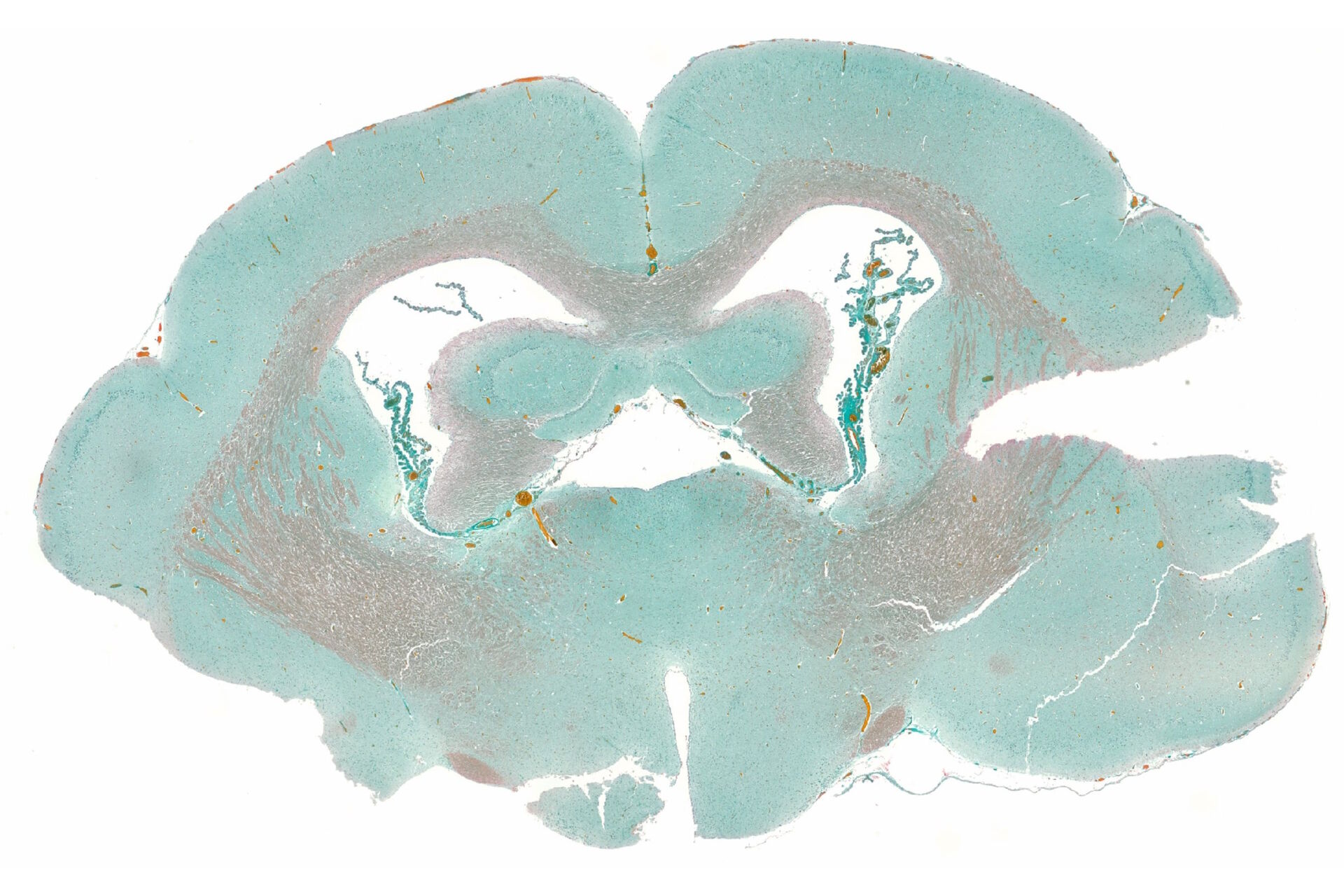
Histological imaging often involvs multiple slides and the need to locate specific regions of interest. Here, we explore how Mica addresses the intricate aspects of histological imaging, ensuring that researchers can work efficiently and with confidence.
One of the initial challenges for histological sample is locating the relevant parts of one or multiple samples. Researchers need to find the sample in 3D, i.e., place the sample above the objective and then focus. But they still need to manually screen the whole sample. Mica's Sample Finder function streamlines this crucial step as it generates an in-focus overview of the slide(s) that can be used for navigational purposes.
Mica's Navigator works like a sophisticated global map composed from satellite images. This intuitive feature allows researchers to zoom in and out on the tissue. It overlays together all captured or selected images for a detailed overview of the tissue sample and to preserve the local context of all acquisitions. The microscope can be efficiently guided to image single positions and large areas.
Imaging large tissue sections requires that sharp focus is always maintained, a demand which is challenging to ensure. Mica combines both hardware and software tools into simple-to-apply focus strategies to help users ensure consistent focus, even for complex imaging scenarios.
To allow examining multiple slides for comprehensive analysis, Mica offers a 4-slide holder. Researchers can efficiently manage and capture images across multiple slides without the need to manually exchange the slide on the holder, enabling higher throughput.

Furthermore, if the contrast is not sufficient, Mica offers IMC, an illumination and detection method to increase contrast based on intrinsic properties of the sample.
Conclusion
For histology, microscopy is indispensable. Histological staining is key to revealing intricate details of cells and tissues. However, choosing between color and fluorescence imaging can be a challenge for researchers. Mica, with its FluoSync technology, provides a one-shot at full sensor resolution solution for both color and fluorescence imaging modes without a compromise. Researchers can have both worlds - high-resolution RGB color and precise fluorescence imaging.
The user-friendly interface, option to handle multiple slides, and features like “sample finder” and context preservation make Mica an ideal choice for scientists performing tissue analysis. This enables researchers to explore cells and tissues rapidly and accurately. Mica is a versatile and powerful solution for researchers working with histologically stained tissue samples. With its innovative FluoSync technology, researchers can analyze histologically stained tissues with ease and confidence.