詳細説明などご希望の場合は、弊社までお問い合わせください。
アプリケーションの分野を選択してください。
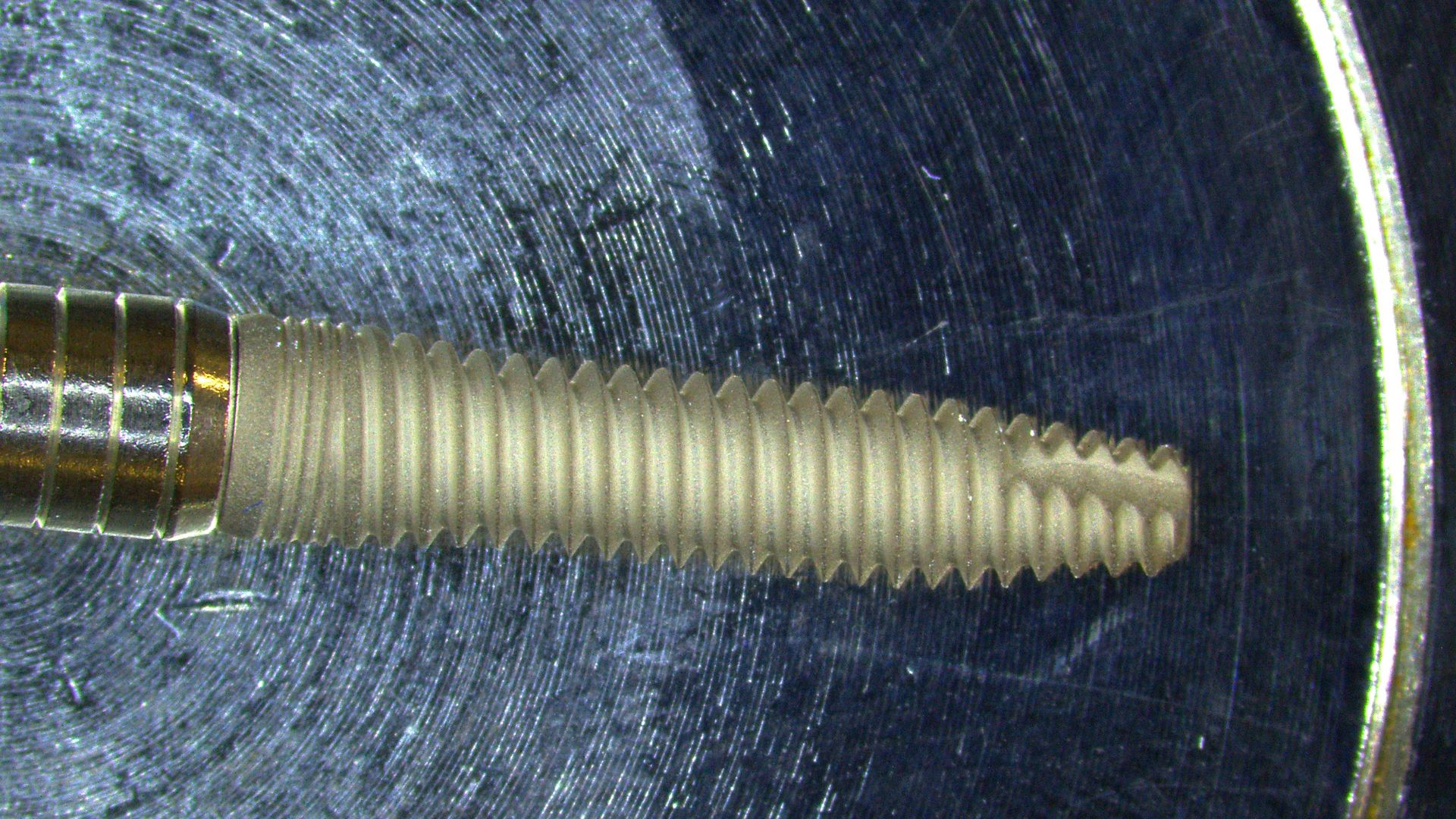

微小な欠陥を確実に検出するには?
適切な解像度と照明を備えた光学顕微鏡を使用することで、医療機器の亀裂やその他の表面の凹凸などの欠陥を正確に検出し、測定することができます。Leicaの実体顕微鏡には、幅広い照明オプションが用意されています。FusionOpticsテクノロジーにより、立体観察で細かい欠陥も見逃さず観察できます。

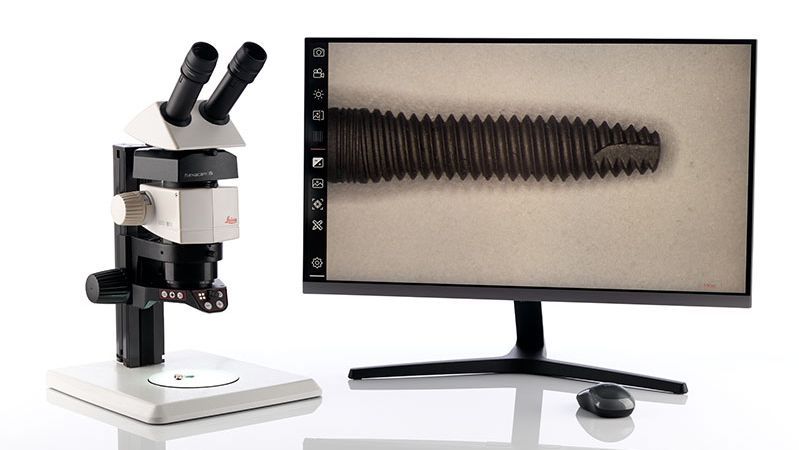
不規則な形状の機器を検査するには?
不規則な形状のデバイスを検査するために深い焦点深度と適切な作動距離を併せ持つ顕微鏡が必要です。最大122mmの作動距離を持つIvesta 3実体顕微鏡で、大きなサンプルや不規則な形状のサンプルの詳細な検査を可能にします。

目視検査中の疲労を避けるには?
人間工学に基づいて設計された顕微鏡ワークステーションを使用することで、作業による疲労や緊張が軽減され、ユーザーの作業環境を改善、集中力を向上させることで、作業の質の向上につながります。Leica実体顕微鏡用の人間工学に基づいたアクセサリーは、長時間の作業でも快適に作業することができます。

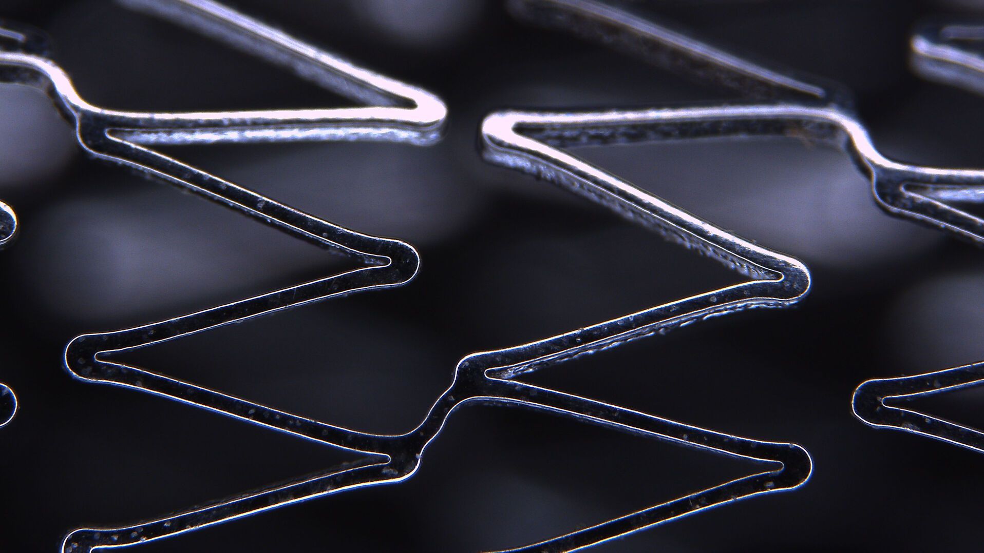
複合材料サンプルの目視検査方法は?
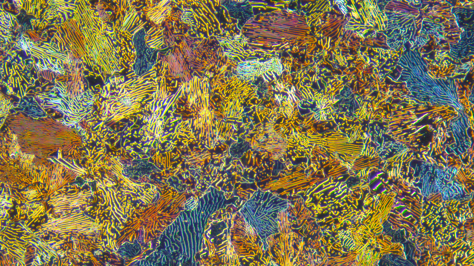
医療機器は複数の材料(金属やプラスチックなど)から構成されることがあります。材料の種類によって、検査時に必要な照明の種類は異なります。Leicaの顕微鏡は幅広い照明オプションを提供し、エッジや表面欠陥を効率的に観察することができます。
Leicaの顕微鏡は、医療機器の品質保証/品質管理の向上にどのように役立っていますか?
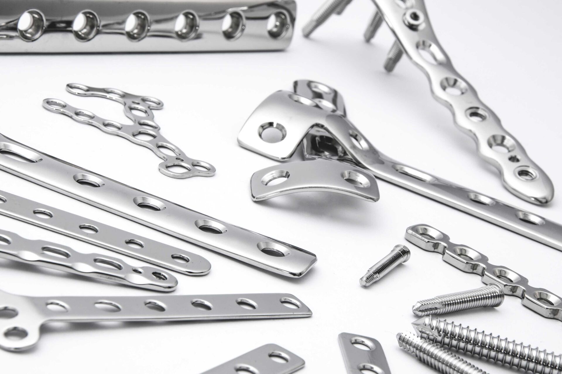
よくある質問 医療機器の品質保証と品質管理 顕微鏡ソリューション
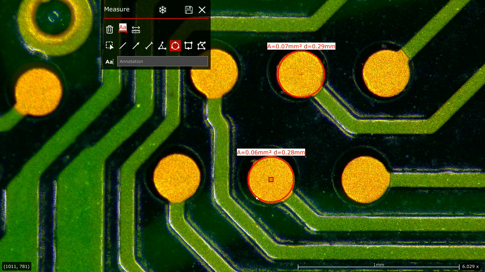
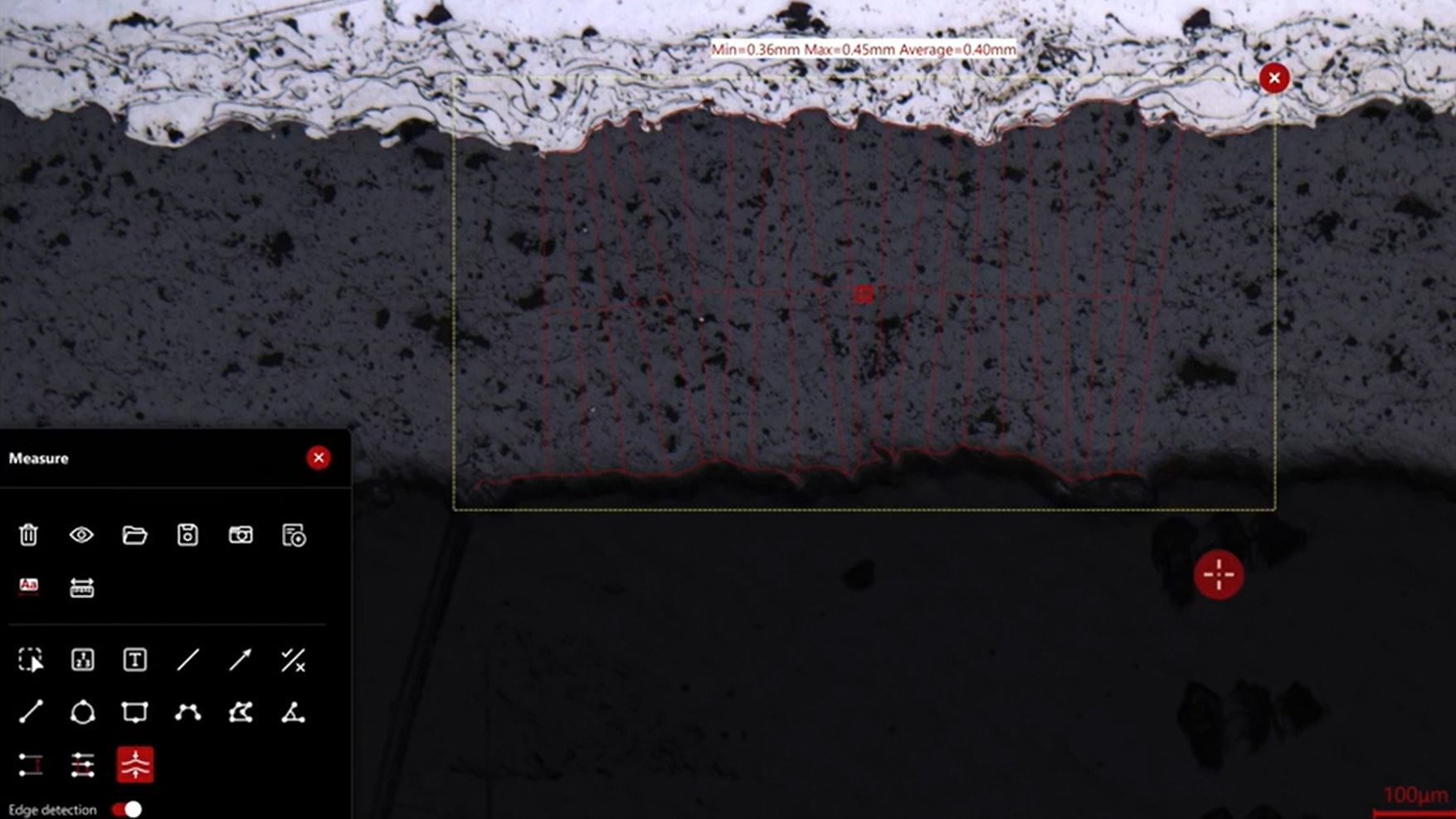
医療機器部品のバリの検出と分析は、光学顕微鏡を使って効率的かつ信頼性の高い方法で行うことができます。バリのサイズは、性能に影響を与えます。顕微鏡による外観検査では、照明とコントラストの方法、解像度、倍率および、画像の撮影時間と保存、画像へのアクセス、使いやすい測定ツールが備わっているか、が重要です。
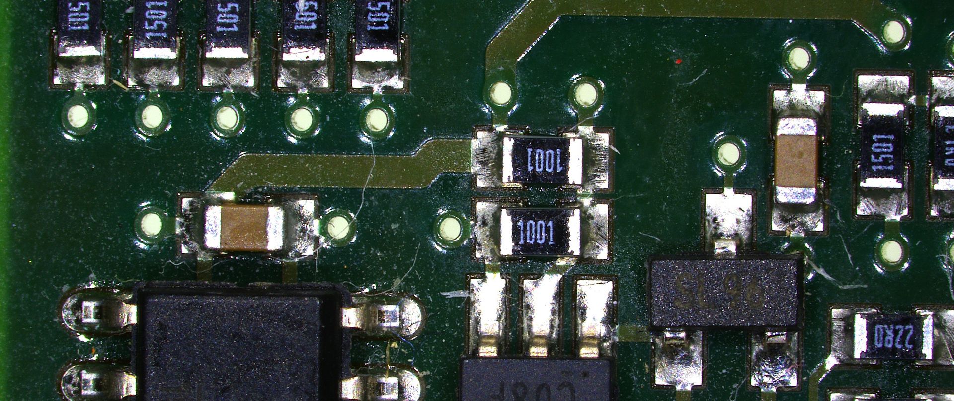
部品検査で必要な光学顕微鏡の倍率と解像度は、対象物の大きさによって異なります。15 µm以上の対象物の場合、最大60倍の倍率と3 µmの解像度が必要です。2µm以上の対象物では、最大約1000倍の倍率が必要で、1µmの解像度に近づきます。0.6µm以上の対象物を観察する場合、最大約1500倍の倍率が必要で、1µm以下の分解能です。
ライカのデジタルマイクロスコープの操作方法は、素早く簡単に習得することができます。一貫性のある直感的なユーザーインターフェイスにより、ユーザーはトレーニングやレビューの時間を短縮できます。ユーザーは顕微鏡の調整にかける時間を最低限にでき、検査により多くの時間を費やすことができます。エンコードされた光学系により、煩雑な作業から解放され、ユーザーは信頼性の高い結果を得ることができます。デジタル画像の表示により、結果を文書化し、他の人と画像共有できます。
ライカの顕微鏡は人間工学に基づいたデザインで、医療機器の外観検査も、操作性を最適化でき、ユーザーの疲労を軽減します。様々なワークフローのニーズへの適応性、生産性の向上、疲労の最小化に重点を置き、ユーザーの要求を念頭に置いて開発されています。人間工学に基づいたデザインは、ユーザーの快適性を保証し、長時間の検査でも負担を軽減します。


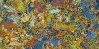
. With DIC users are able to visualize small height differences on the wafer surface more easily.")

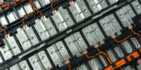
 at the edge of a battery electrode acquired with a DVM6 digital microscope.")