
正確な結果
難しいと言われている偏光観察は、シンプルなステップで最適な結果を得ることが重要です。
- 光学歪のない光学系を採用し、鮮明な観察像
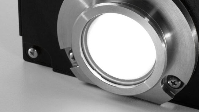
- 最新のLED照明により、明るく照明ムラのない、一定の色温度を提供
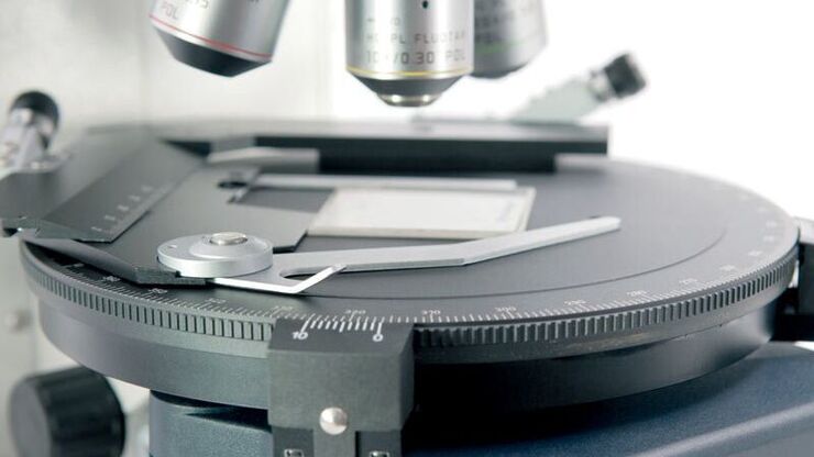
- 回転心出し機構付回転ステージによる、効率的な偏光観察
- ベルトラン絞りの採用により、シャープなコノスコープ像が観察
偏光顕微鏡の詳細はScience Labを参照ください。 polarization microscopy on Science Lab.
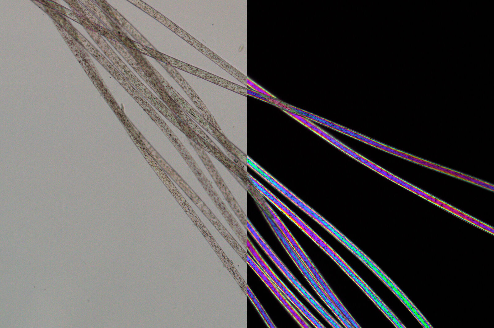
落射・透過照明
ライカ偏光顕微鏡は、透過光あるいは落射光どちらかにLED照明を搭載、あるいは透過光と落射光両方にLED照明を構成することも可能です。
- 落射照明: 岩石、鉱物の反射率測定など
- 透過照明: 金属や不透明な結晶、ポリマー、医薬品の観察、複屈折率測定など
- 落射および透過照明: 地質研究など
マクロからミクロまで、ダイナミックな観察
芯出し可能な6 穴対物レボルバ付、ニーズにあわせて最適な倍率で拡大観察できます。
- 2.5x対物レンズ: サンプルの全体像を観察
- 63x対物レンズ: シャープなコノスコープ像
- 100×対物レンズ: 粒度と結晶粒の分布解析
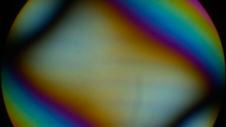
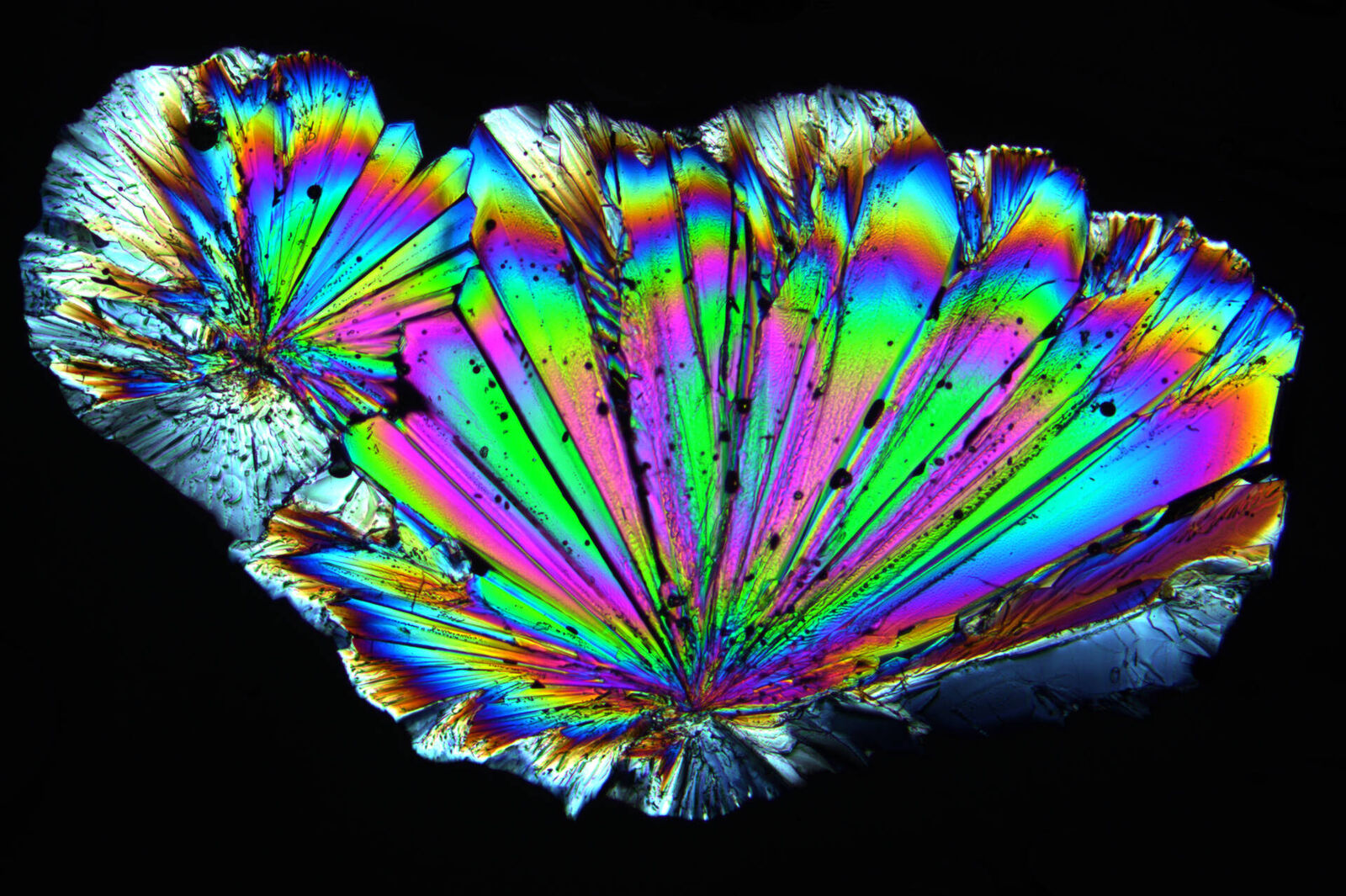
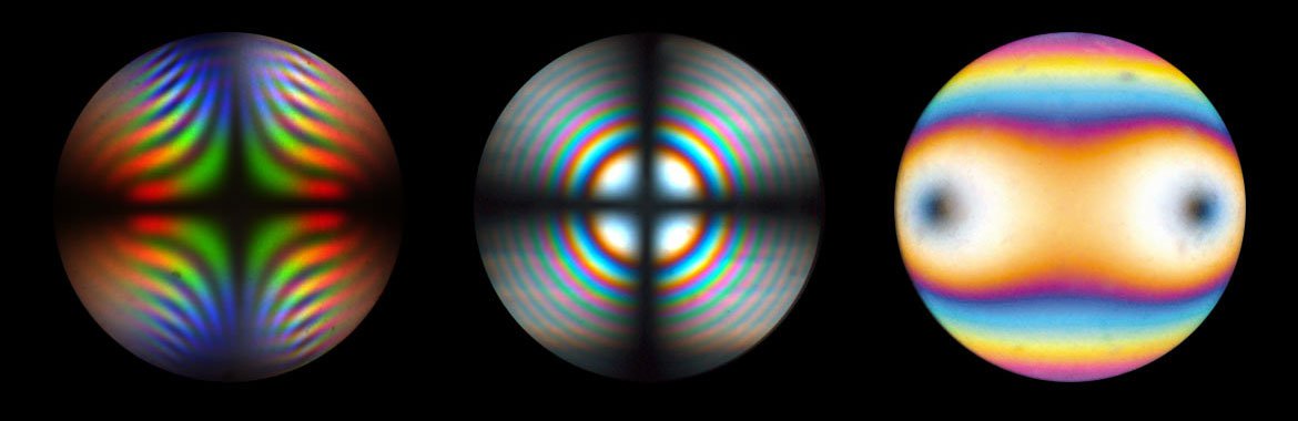
コノスコープ観察
コノスコープ観察は、一軸、二軸性の判定や光軸角の測定等、岩石鉱物をはじめとする結晶を同定するのに使用します。
Bi-axial interference figure of thin biotite crystal in diagonal position at circular polarized light. Position of optical axis can be clearly identified
Leica DM4 Pコノスコープ観察 :
- 光学歪を極限まで除去した光学系の採用で、鮮明な偏光観察
- 充実したアクセサリとの組み合わせで高度な偏光観察、測定
Images recorded with a DM4 P microscope using transmitted light, conoscopy, 63x N Plan objective, and polarizers


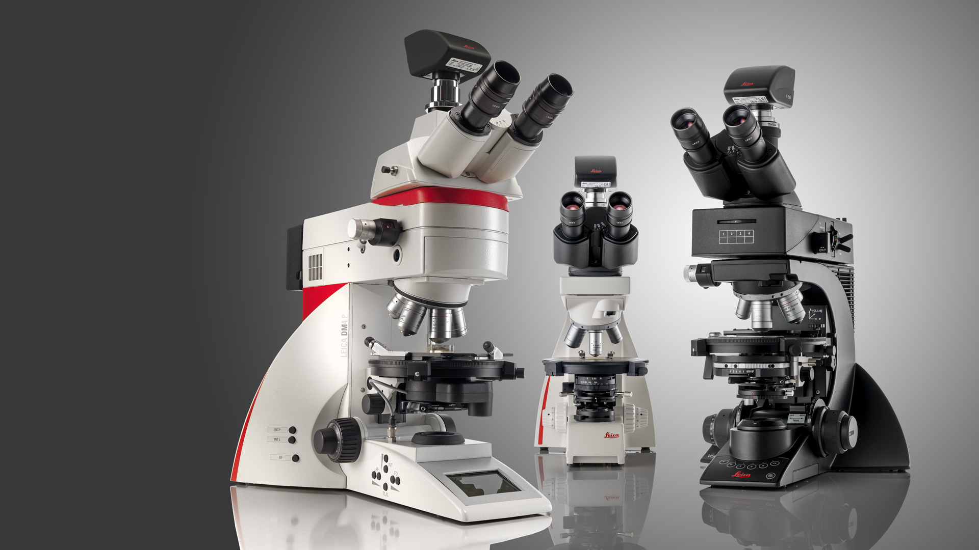
3つのモデルから最適な機種を選択いただけます
アプリケーションに応じて、3つのモデルから最適な機種を選択いただけます。
- ライカ DM4 P:電動制御モデル
- ライカ DM2700 P:マニュアルモデル
- ライカ DM750 P:大学その他における教育向け
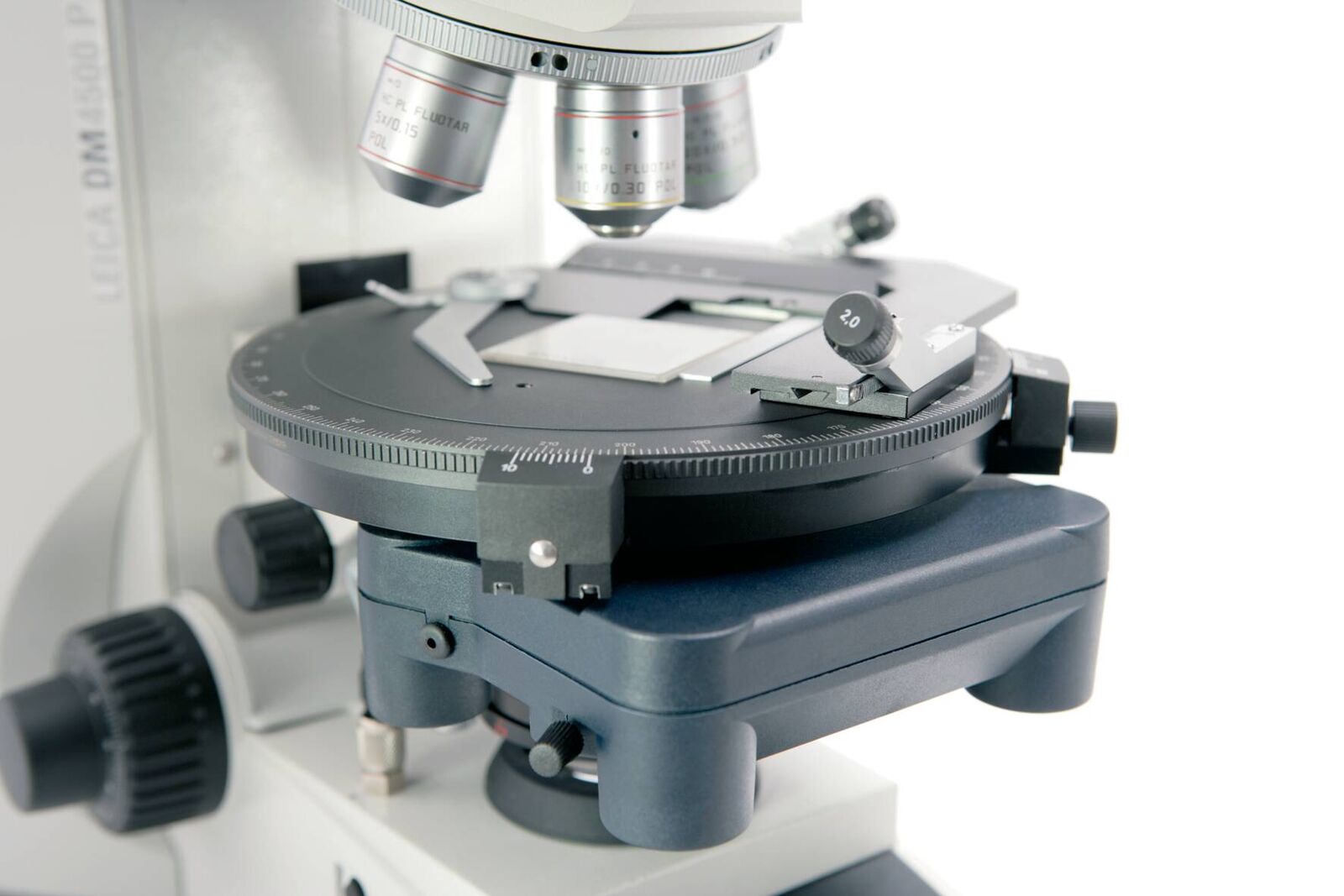
高性能・電動制御モデル: Leica DM4 P
フル・セミ電動制御タイプの、システム偏光顕微鏡です。オルソスコープ、コノスコープ観察に対応、豊富なアクセサリとの組み合わせで高度な偏光観察、測定が可能です。
- 対物レンズ情報読取り(コーディング)、センタリング式 6 穴対物レボルバ
- 倍率を変更しても明るさ、コントラストを自動調整
- 最新の LED 技術で、視野周辺まで照明ムラなく、一定の色温度
- 顕微鏡条件がすぐわかる液晶ディスプレイ
- 45°クリックストップ(オプション)、高精度360°回転ステージ
- 光学歪のない高い光学性能
- 高度な偏光性能を提供する、豊富なアクセサリ
- DIN 58879対応コンペンセータ
研究用途からルーティン検査まで: ライカ Visoria P
コード化された機能、最適化されたイルミネーションマネージャーなどの機能により、ワークフローを効率化します。顕微鏡の人間工学に基づいた設計により、より快適に、身体負担を最小限に抑えることができます。
- コード機能付 5穴センタリング対物レボルバ
- UC-3D照明
- イルミネーションマネージャーで再現性の高い結果
- カラーコードされた絞りのアシスタント機能
- フォーカスストップ内蔵
- 3ギアフォーカスドライブ
- LED照明
- 幅広い種類のコノスコピーモジュール
- 歪のない光学系
- 360°回転ステージ(45°クリックストップ付き・なし)
- 豊富な偏光装置
- DIN 58879に準拠した固定および可変コンペンセーター
エントリー・教育実習用に最適: Leica DM750 P
エントリーモデルながら、高い光学性能を提供し、本格的な偏光観察が可能
- 可搬性も考慮された優れたデザイン、ケーブルもコンパクトに収納可能
- 2つのコンペンセータ収納可能
- 178mmと広い回転ステージ
- センタリング式4穴対物レボルバ
