STELLARIS CRS
共焦点顕微鏡
製品紹介
Home
Leica Microsystems
STELLARIS CRS コヒーレントラマン散乱顕微鏡
蛍光標識不要の化学顕微鏡
最新の記事を読む
 dataset, showing the biochemically distinct structures of a fresh, untreated apple slice.")
How to Prepare Samples for Stimulated Raman Scattering (SRS) imaging
Find here guidelines for how to prepare samples for stimulated Raman scattering (SRS), acquire images, analyze data, and develop suitable workflows. SRS spectroscopic imaging is also known as SRS…
, unsaturated lipids (magenta, 3050 cm-1), collagen (SHG, cyan). Sample courtesy of R. Rudolf, J Klicks, Hochschule Mannheim")
The Potential of Coherent Raman Scattering Microscopy at a Glance
Coherent Raman scattering microscopy (CRS) is a powerful approach for label-free, chemically specific imaging. It is based on the characteristic intrinsic vibrational contrast of molecules in the…

Formulated Product Characterization with SRS Microscopy
Creams, pastes, gels, emulsions, and tablets are ubiquitous across a wide range of manufacturing sectors from pharmaceuticals and consumer health products to agrochemicals and paint. To improve…

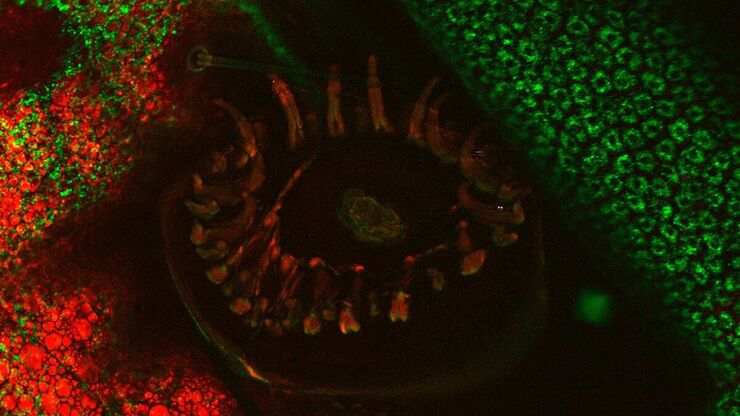
研究におけるモデル生物
モデル生物とは、特定の生物学的プロセスを研究するために研究者が使用する生物種です。 モデル生物は、人間と似た遺伝的特徴を持ち、遺伝子学、発生生物学、神経科学などの研究分野で一般的に使用されています。 通常、モデル生物は実験環境での維持や繁殖が容易であること、生殖サイクルが短いこと、または、特定の形質や病気を研究するために突然変異体を生成する能力を持つことで選ばれます。

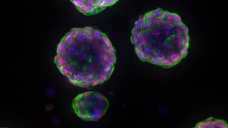
がん研究
がんは、成長調節における欠損細胞によって引き起こされる複雑な異質性疾患です。 細胞または細胞群内の遺伝的および後成的変化が通常の機能を妨げ、自律的、非制御の細胞成長と増殖を引き起こします。
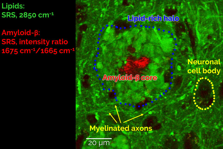
Stimulated Raman Scattering Microscopy Probes Neurodegenerative Disease
Despite decades of research, the molecular mechanisms underlying some of the most severe neurodegenerative diseases, such as Alzheimer’s or Parkinson’s, remain poorly understood. The progression of…

CARS Microscopy: Imaging Characteristic Vibrational Contrast of Molecules
Coherent anti-Stokes Raman scattering (CARS) microscopy is a technique that generates images based on the vibrational signatures of molecules. This imaging methods does not require labeling, yet…
An Introduction to CARS Microscopy
CARS overcomes the drawbacks of conventional staining methods by the intrinsic characteristics of the method. CARS does not require labeling because it is highly specific to molecular compounds which…
バイオファーマ
ライカのソリューションは、バイオ医薬品業界において、創薬の促進、細胞解析の強化、規制を満たすデータの完全性をサポートします。
Fields of Application
バイオファーマ
ライカのソリューションは、バイオ医薬品業界において、創薬の促進、細胞解析の強化、規制を満たすデータの完全性をサポートします。
