Specialità mediche
Specialità mediche
Esplora una raccolta completa di risorse scientifiche e cliniche su misura per gli operatori sanitari, tra cui approfondimenti tra pari, casi clinici e simposi. Progettata per neurochirurghi, oculisti e specialisti in chirurgia plastica e ricostruttiva, otorinolaringoiatria e odontoiatria. Questa raccolta evidenzia gli ultimi progressi nel campo della microscopia chirurgica. Scoprite come le tecnologie chirurgiche all'avanguardia, come la fluorescenza AR, la visualizzazione 3D e l'imaging OCT intraoperatorio, consentano di prendere decisioni sicure e di essere precisi in interventi chirurgici complessi.
Consumables for Laser Microdissection
There are many different types of consumables for laser microdissection (LMD) systems. They cover a wide range of applications from basic to highly specialized, enabling scientists to choose their own…
How FLIM Microscopy Helps to Detect Microplastic Pollution
The use of autofluorescence in biological samples is a widely used method to gain detailed knowledge about systems or organisms. This property is not only found in biological systems, but also…
High-pressure freezing: Revealing functional mechanisms of synaptic transmission
Learn more about applying optogenetic stimulation in the EM ICE and how this technology has the potential to reveal structural and functional mechanisms of synaptic transmission. Get a detailed…
Workflows and Instrumentation for Cryo-electron Microscopy
Cryo-electron microscopy is an increasingly popular modality to study the structures of macromolecular complexes and has enabled numerous new insights in cell biology. In recent years, cryo-electron…
Organismi Modello nella Ricerca
Un organismo modello è una specie utilizzata dai ricercatori per studiare specifici processi biologici. Hanno caratteristiche genetiche simili a quelle umane e vengono normalmente impiegate in aree di…
An Introduction to Computational Clearing
Many software packages include background subtraction algorithms to enhance the contrast of features in the image by reducing background noise. The most common methods used to remove background noise…
How Can Immunofluorescence Aid Virology Research?
Modern virology research has become as crucial now as ever before due to the global COVID-19 pandemic. There are many powerful technologies and assays that virologists can apply to their research into…
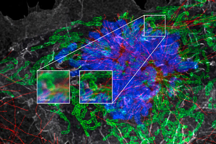
Utilizza LIGHTNING per ottenere il massimo delle informazioni dal tuo campione
LIGHTNING è un processo di estrazione di informazioni completamente automatico, che funziona in modo adattivo, grazie al quale si possono apprezzare strutture e dettagli fini dei campioni altrimenti…
Virologia
L’oggetto della tua ricerca si concentra su infezioni e malattie virali? Scopri in che modo puoi approfondire l’aspetto virologico utilizzando le soluzioni di imaging e la preparazione del campione di…
