Murine esophageal organoids (DAPI, Integrin26-AF 488, SOX2-AF568) imaged with THUNDER Imager Cell. Courtesy of Dr. F.T. Arroso Martins, Tamere University, Finland.

バイオ医薬品向け先進イメージング・ソリューション

高度なイメージング・ソリューションは、創薬、品質管理、複雑な生物学的システムの理解において、バイオ医薬品研究者に重要な知見を提供します。ライカの顕微鏡ソリューションは、イノベーション、ユーザー中心の設計、信頼性の高いデータインテグリティを融合し、ブレークスルーと一貫した研究成果をサポートします。

お気軽にご連絡ください!

ライカの画像処理の専門家が、バイオ医薬品業界向けのソリューションについてアドバイスいたします。

Co-culture of CAR-T cells and breast cancer organoids. CAR-T cells labeled by CD3 (green). Breast cancer organoids labeled by membrane marker B7H3 (blue). DNA labeled by SiR-DNA (red). Image acquired using STELLARIS. Image courtesy of Dr Ravian van Ineveld and Prof. Dr Anne Rios, DREAM3D Lab.

3D細胞培養を効果的に画像化するには?

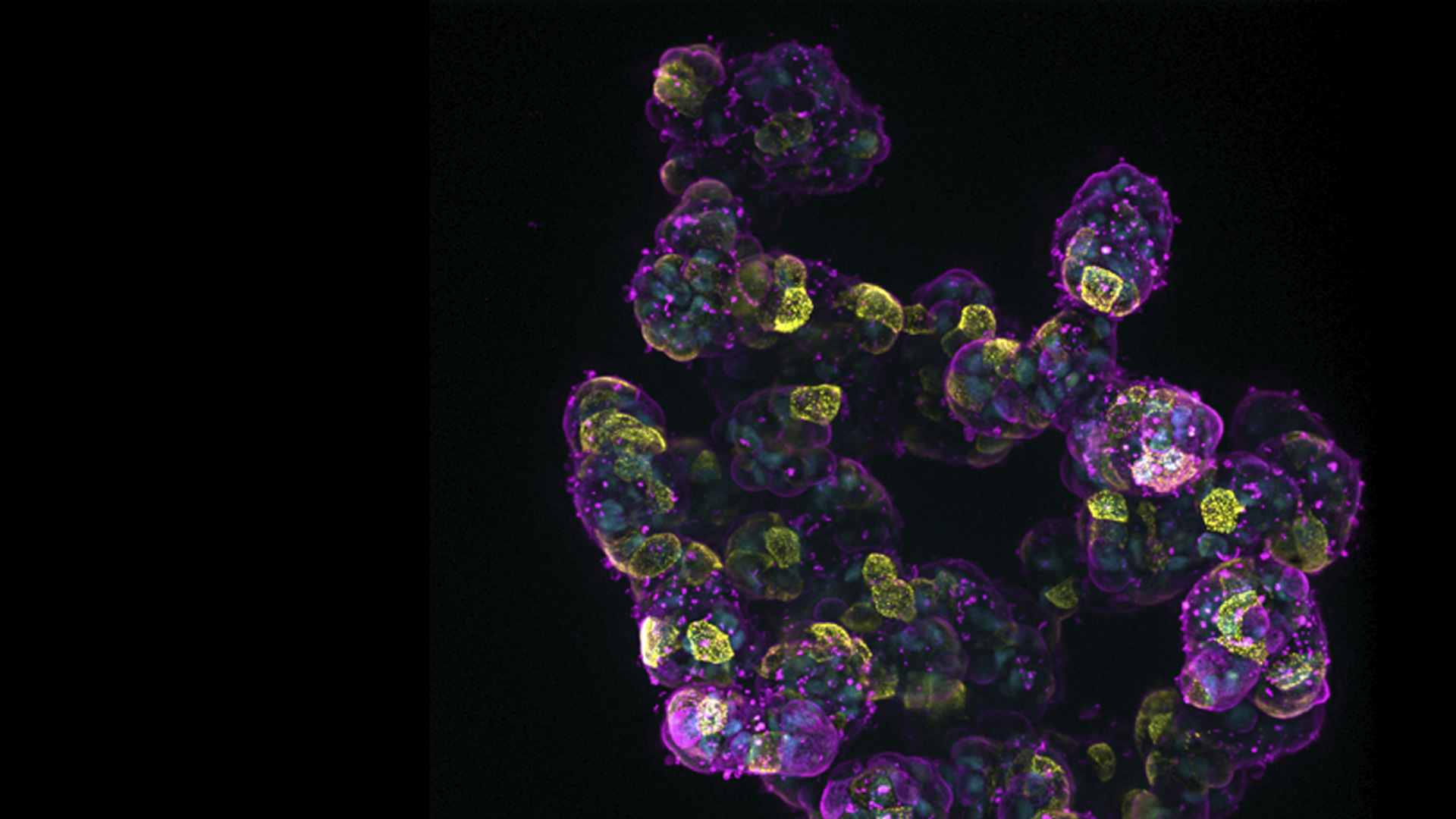
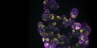
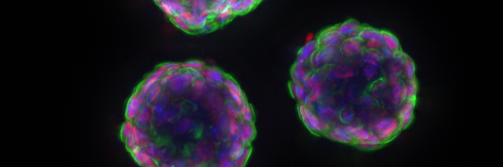
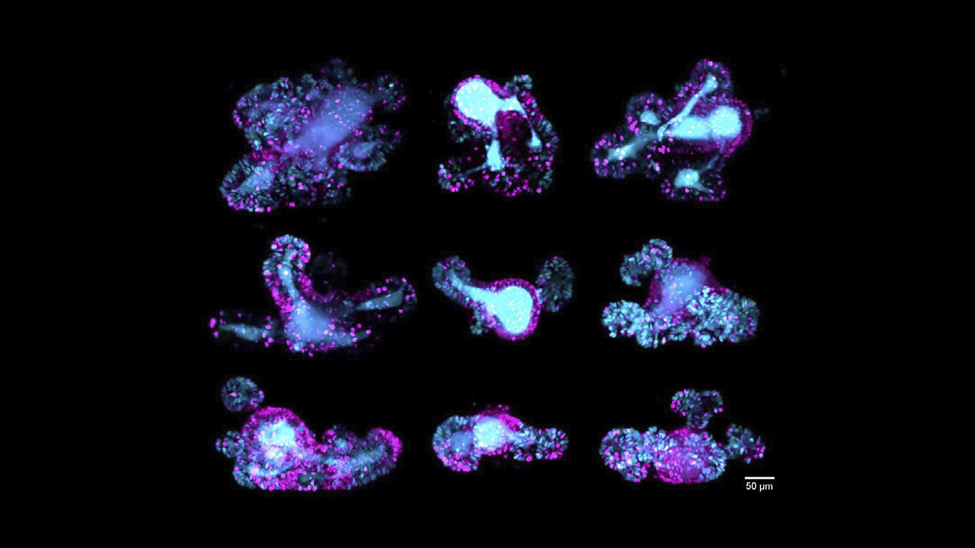
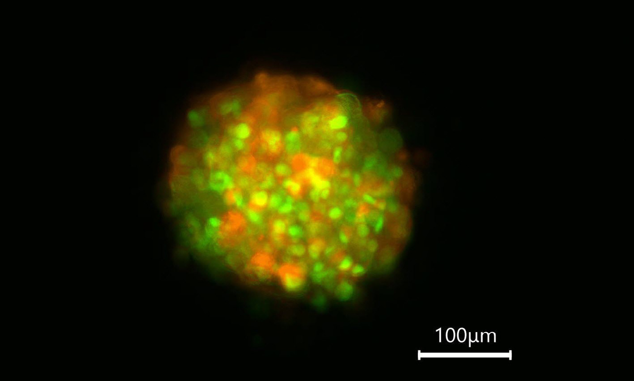
ライカの先進的なイメージング・ソリューションを使用することで、画像データに含まれていた詳細情報を解き放ちます。例としては、細胞の深部をイメージングする際の焦点外のぼけが挙げられます。STELLARISコンフォーカル・プラットフォームやViventis Deep ライトシート イメージングシステムのような光学切片化技術は、通常では目に見えない微細な構造や詳細を明らかにする洞察の抽出に役立ちます。

細胞増殖/遊走を正確に定量化するには?

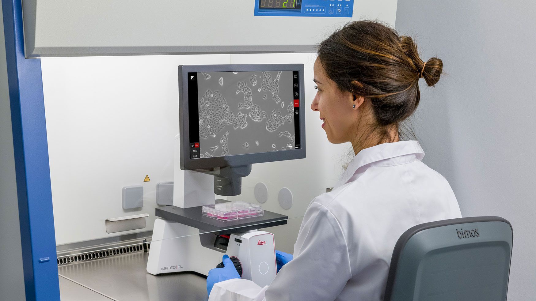
ライカのシステムは、生細胞の遊走、増殖、分化を正確に定量化し、エンドポイント研究を可能にします。Aiviaの機械学習アルゴリズムのサポートにより、バイオファーマの科学者は、このような科学的な質問に関連する一般的な手作業の複雑さを克服することができます。

どうすれば空間生物学を始めることができますか?

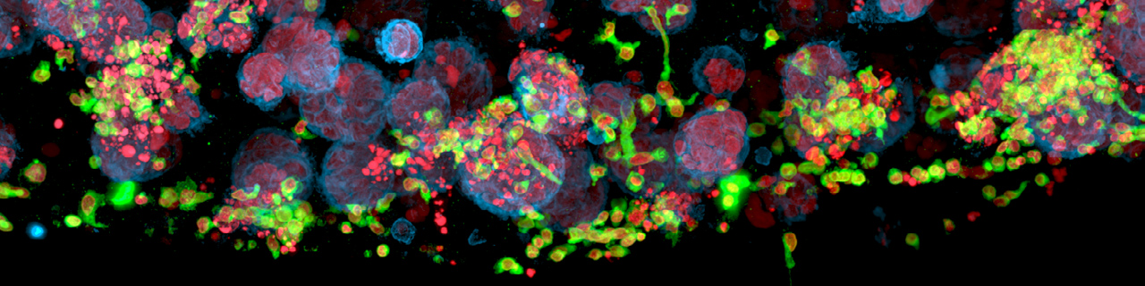
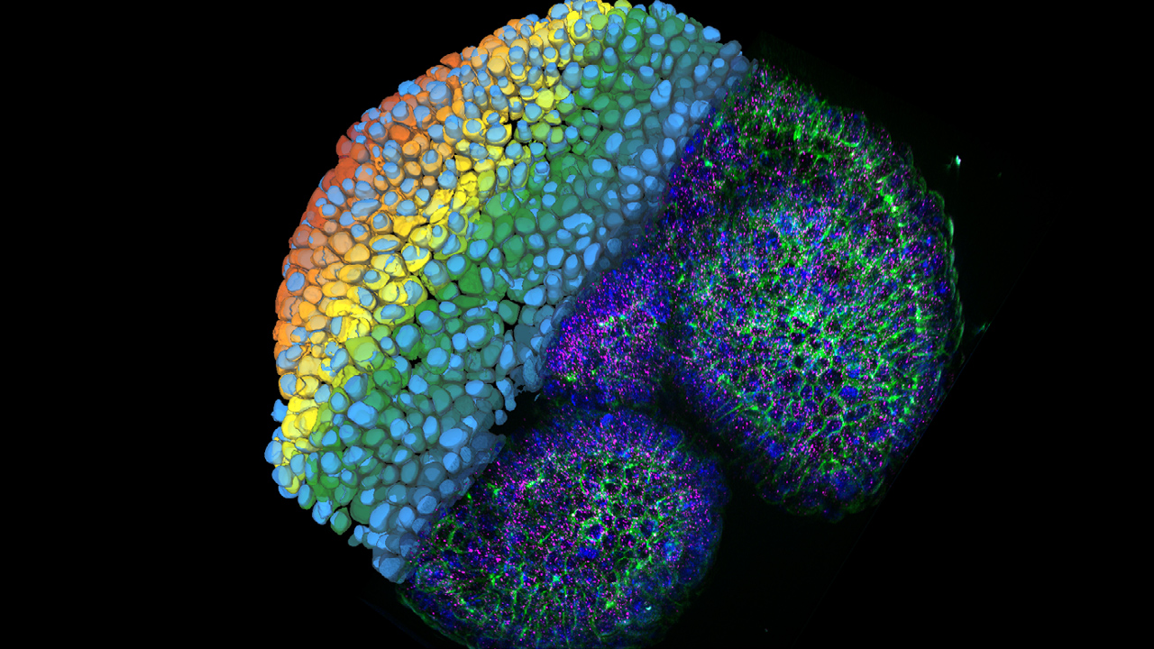
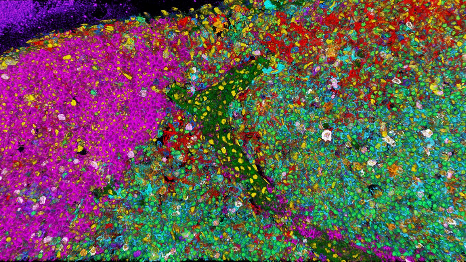
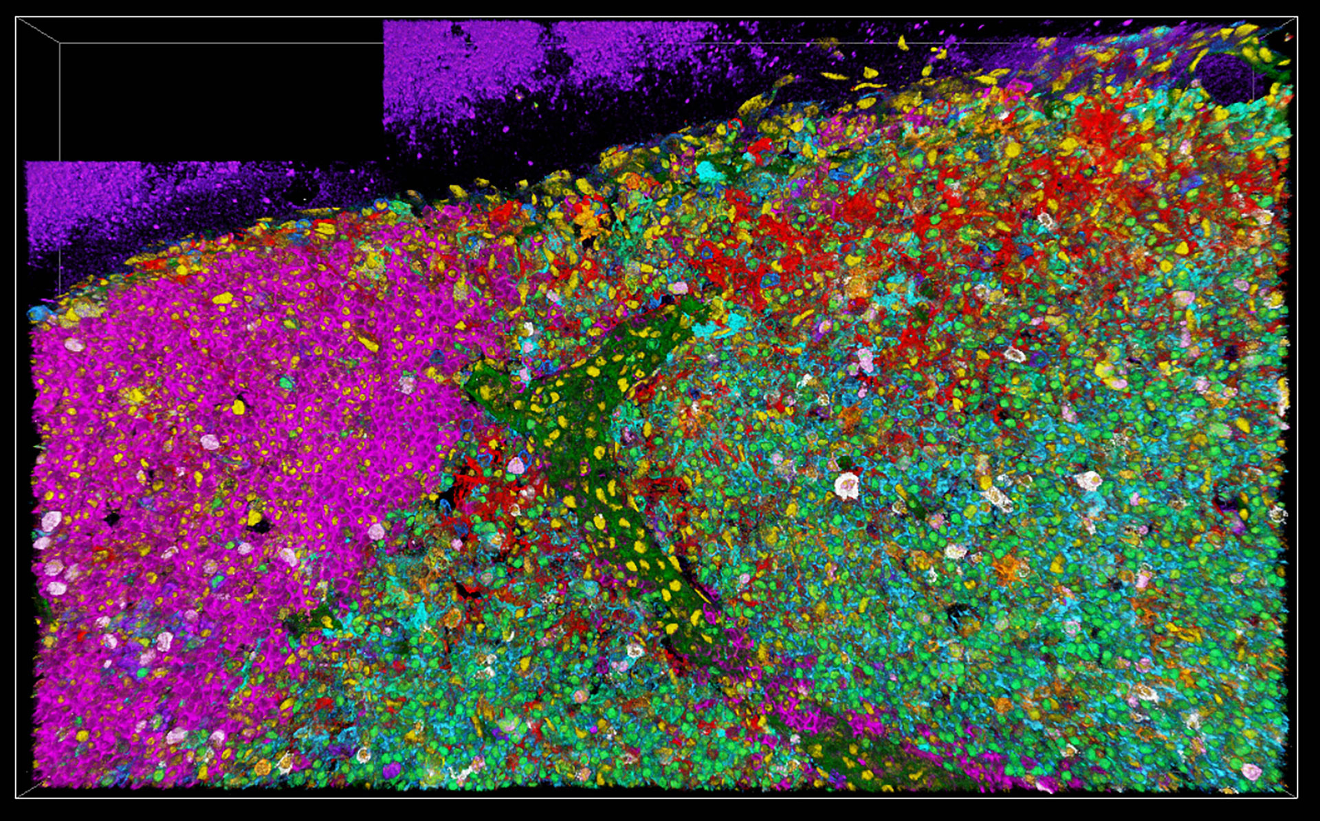
SpectraPlex for STELLARISにより、バイオ医薬品研究者は、15以上のバイオマーカーを含む空間生物学用の複雑な標本を一度に複数のスケールおよび3Dで画像化することができます。Cell DIVEは、反復多重化のための抗体使用に関するガイドラインを、テスト済みのデータベースとともに提供します。さらに、Aiviaは空間生物学的解析を強化し、標準化された使いやすいプロトコルに統合しています。

空間イメージングでメカニズムを検証できますか?

はい、蛍光寿命イメージング、超解像、多光子顕微鏡、ライトシートおよびラベルフリーの化学イメージング、細胞および組織の空間発見のための3Dハイプレックスイメージングを利用することで可能です。STELLARIS共焦点プラットフォームは、空間生物学に関連するメカニズムの検証に役立つこれらのマルチモーダル機能を提供します。

ライカの先進的な顕微鏡ソリューションは、バイオ医薬品研究者にとってどのような利点がありますか?


信頼できるデータを。品質管理のためのCFR準拠蛍光イメージング

Mateo FLは、FDA 21 CFR Part 11コンプライアンスをサポートする監査証跡とユーザー管理機能を備えており、バイオ医薬品のワークフローにおけるデータ管理をより簡単かつ安全にします。


正確な分析のためのラベルフリー分子イメージング

STELLARIS CRSは、ラベルフリーの分子イメージングを提供し、バイオ医薬品研究者がサンプルの完全性を損なうことなく、複雑なサンプルを分析するという課題を克服することを可能にします。高速で非破壊的なイメージングにより、研究者が創薬、製剤化、疾患研究において分子解析に取り組むのに役立ちます。


3Dサンプルの高品質データ抽出と長時間のボリュームイメージング

ハイスループット・スピニングディスク・コンフォーカルにより、複雑な3Dサンプルからより統計的に有意なデータを短時間で抽出します。Viventis Deepは、複数のオルガノイドのサンプル体積を最大2倍まで、時間経過に伴う前例のない詳細さで並行して穏やかに可視化する手段を提供します。

初期の研究:病気のメカニズムを理解する

初期段階のバイオ医薬品研究は、疾患の細胞および分子メカニズムを解明することに焦点を当てています。生物学的構造や相互作用を本来の文脈で可視化するためには、高解像イメージングが不可欠です。

ライカの先進的な共焦点顕微鏡、超解像顕微鏡、ワイドフィールド顕微鏡は、細胞の精密なイメージングを可能にします。ライブセルイメージングやタイムラプス研究をサポートし、細胞の挙動と疾患の進行に関する重要な洞察を提供します。直感的なライカのソフトウェアとAIによる画像解析により、研究者はデータの解釈と仮説の立案を加速することができます。

3D 15-plex imaging for phenotyping in immune response. Image acquired using STELLARIS with SpectraPlex. Roberti J., Hecht F., Gai E., Straka T., Holzmeister S., Steinmetz I., Wong H., Alvarez L. Nat. Methods (2024).

創薬:ターゲットの特定と検証

創薬において、生物学的標的の同定と検証は極めて重要です。タンパク質の発現、バイオマーカーの分布、細胞応答を可視化することは、このプロセスにおいて重要な役割を果たします。研究者は、免疫蛍光やタンパク質の局在研究をサポートする最先端のイメージングツールを必要としています。

ライカの共焦点顕微鏡と超解像顕微鏡は、大規模なサンプルセットのスクリーニングや分析に必要な鮮明さとスピードを提供します。自動化機能により画像取得と解析が合理化され、再現性が可能になり、人的ミスが減少します。

医薬品製造& 品質管理:一貫性とコンプライアンスの確保

製造および品質管理(QC)環境では、製品の完全性、無菌性、規制遵守を維持することが必須となります。目視検査は、各工程を検証する上で重要な役割を果たします。

Mateo FLデジタル蛍光顕微鏡は、マルチモード蛍光・透過光撮影、自動解析ツール、安全なデータトラッキングにより、高度な細胞培養研究を支援します。FDA 21 CFR Part 11に準拠した監査証跡とユーザー管理機能を有しており、より簡単で安全なデータ管理が可能です。

Brain organoids labeled with lamin (green) and tubulin (magenta), acquired using Viventis Deep Dual View Light Sheet Microscope. Courtesy of Akanksha Jain, Treutlein Lab ETH-DBSSE Basel (Switzerland).

医薬品開発:前臨床およびトランスレーショナル・リサーチ

医薬品候補が開発段階に進むにつれ、厳格な試験と深い生物学的洞察が不可欠になります。イメージングは、研究者が有効性、安全性、作用機序を評価するのに役立ちます。

ライカマイクロシステムズのTHUNDER Imager Cell Spinning Diskは、研究者に3Dサンプルの高品質データと鮮明なディテールを提供し、研究をより深く、より詳細に進めることができます。

バイオファーマでよくある質問

関連記事

バイオ医薬品向けイメージング・ソリューションに関する最新記事を読む

ライカ マイクロシステムズの知識ポータルでは、顕微鏡に関する科学的な研究記事および教材を提供しています。日常業務や研究で、ビギナーの方から経験豊富な方まで幅広くサポートします。

その他の記事
Brain organoid labeled with lamin (green) 
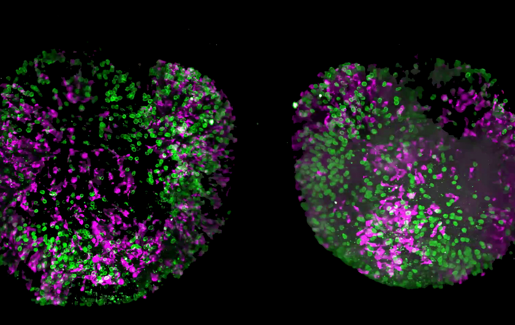
and tubulin (magenta), acquired using Viventis Deep. Courtesy of Akanksha Jain, Treutlein Lab ETH-DBSSE Basel (Switzerland).

Faster & Deeper Insights into Organoid and Spheroid Models

Gain deeper, more translatable, insights into organoid and spheroid models for drug discovery and disease research by overcoming key imaging challenges. In this eBook, explore advanced microscopy…
Pancreatic Ductal Adenocarcinoma with 11 Apoptosis biomarkers shown – BAK, BAX, BCL2, BCLXL, Caspase9, CIAP1, NaKATPase, PCK26, SMAC, Vimentin, and XIAP.

Transforming Research with Spatial Proteomics Workflows

Spatial Proteomics, Nature Methods 2024 Method of the Year, is driving research advancements in cancer, immunology, and beyond. By combining positional data with high throughput imaging of proteins in…
Digital microscopy simplifies documenting cell-culture results electronically while following 21 CFR part 11 guidelines for biopharma.

Introduction to 21 CFR Part 11 for Electronic Records of Cell Culture

This article provides an introduction to the recommendations of 21 CFR Part 11 from the FDA, specifically focusing on the audit trail and user management in the context of cell-culture laboratories.…
Automated Laser Microdissection for Proteome Analysis

Deep Visual Proteomics Provides Precise Spatial Proteomic Information

Despite the availability of imaging methods and mass spectroscopy for spatial proteomics, a key challenge that remains is correlating images with single-cell resolution to protein-abundance…
AI-based transfection analysis (left) 
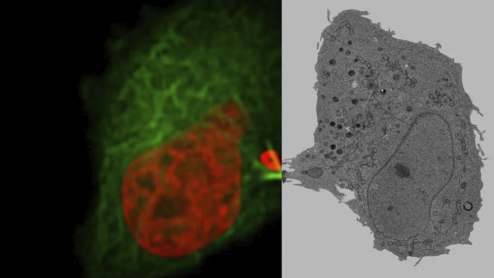
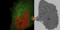
of U2OS cells which were transfected with a fluorescently labelled protein. A fluorescence image of the cells (right) is also shown. The analysis and imaging were performed with Mateo FL.

Leveraging AI for Efficient Analysis of Cell Transfection

This article explores the pivotal role of artificial intelligence (AI) in optimizing transfection efficiency measurements within the context of 2D cell culture studies. Precise and reliable…
Living HeLa cells stained with WGA-488 (yellow)
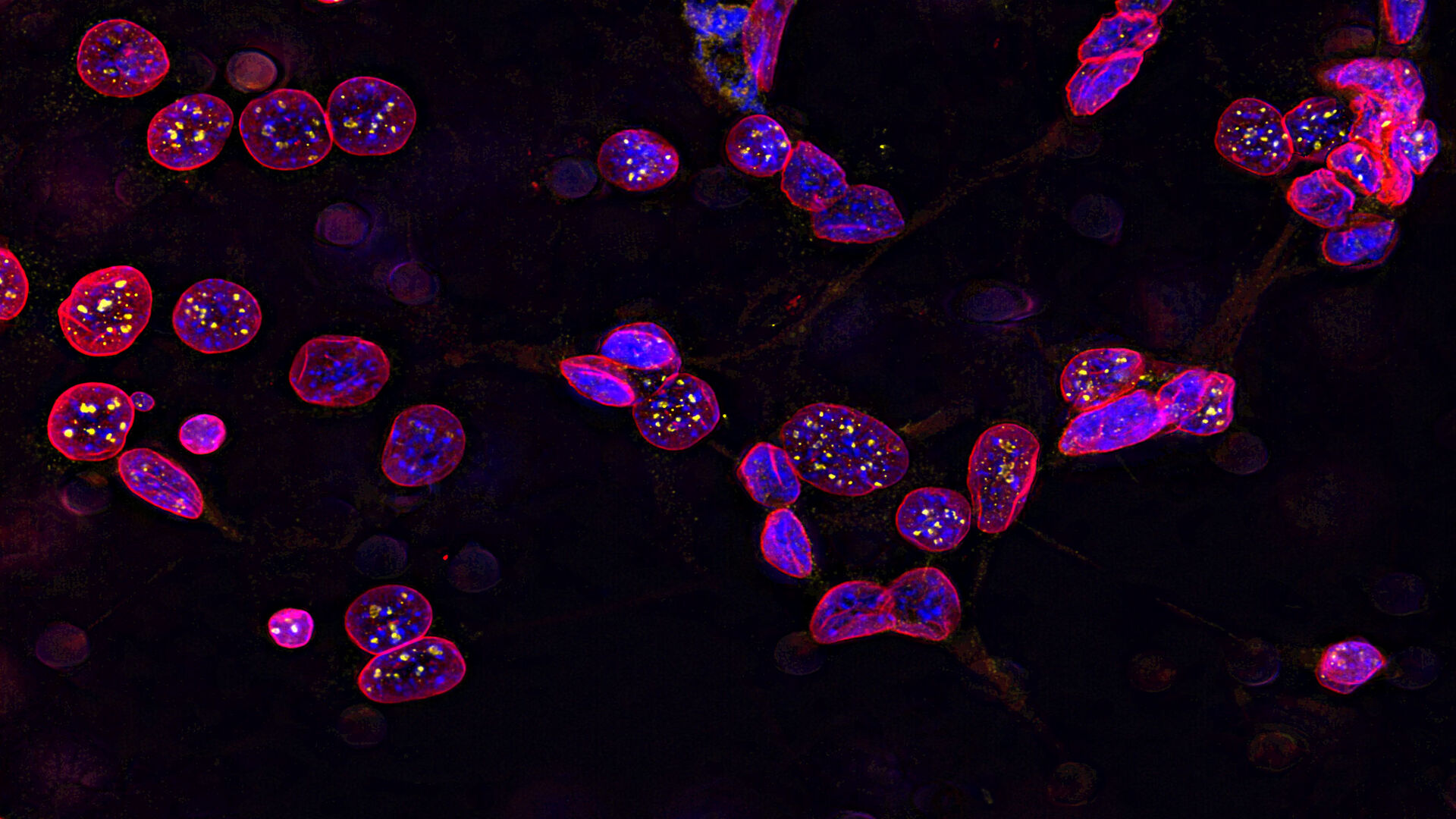
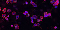
, SPY-Actin (cyan), and SiR-Tubulin (magenta). Instant Computational Clearing (ICC) was applied.

How to Perform Dynamic Multicolor Time-Lapse Imaging

Live-cell imaging sheds light on diverse cellular events. As many of these events have fast dynamics, the microscope imaging system must be fast enough to record every detail. One major advantage of…
Virally labeled neurons (red) and astrocytes (green) in a cortical spheroid derived from human induced pluripotent stem cells. THUNDER Model Organism Imager with a 2x 0.15 NA objective at 3.4x zoom was used to produce this 425 µm Z-stack (26 positions), which is presented here as an Extended Depth of Field (EDoF) projection.  Images courtesy of Dr. Fikri Birey  from the Dr. Sergiu Pasca laboratory at Stanford University, 3165 Porter Dr., Palo Alto, CA

Download The Guide to Live Cell Imaging

In life science research, live cell imaging is an indispensable tool to visualize cells in a state as in vivo as possible. This E-book reviews a wide range of important considerations to take to…
14: 7-day old 3D cell culture of MDCK cells stably transfected with; Mx1-GFP (green), SPY555-Actin (yellow) and WGA-AlexaTM 647 (magenta), and imaged using a THUNDER Imager Cell Spinning Disk. Shown are both the spinning disk image (left) and the same image after THUNDER Computational Clearing was applied (right); scale bar 50 μm.

オルガノイドと3D細胞培養

ライフサイエンス研究で近年最も目覚ましい発展の一つは、オルガノイド、スフェロイド、生体機能チップモデルなどの3D細胞培養システムの開発です。 3D細胞培養は、細胞が成長し、全3次元で周囲と相互作用できる人工環境です。 これらの条件は、生体内条件に似ています。

もっと読む

Image of C2C12 cells: The cells are stained with lamin B (magenta) which indicates nuclear structure, Hoechst (blue) indicating DNA, and γH2AX (yellow) indicating damage to DNA. Cells were imaged using a THUNDER Imager 3D Live Cell with a 63X/1.4 oil immersion objective.

細胞生物学研究

細胞生物学イメージングは、さまざまな光電子相関顕微鏡を使用して行われます。 ライカマイクロシステムズが提供するイメージングソリューションは、細胞生物学研究を最大限に行えるように設計されています。

もっと読む

Mammalian cells expressing H2B-mCherry and alphaTubulin-mEGFP to visualize DNA and microtubules respectively. Left: Widefield imaging using THUNDER Imager. Right: Sample after correlated EM imaging using Leica Microsystems Coral Life workflow. Cells were cryo-immobilized by high-pressure freezing 60 min after the onset of cytokinesis. The intercellular bridge was imaged under TEM after freeze substitution and resin infiltration.

光電子相関顕微鏡法(CLEM)

ライカマイクロシステムズ独自のCLEMソリューションは、サンプルの生存性や品質チェック、精密かつ確実な3Dターゲティング機能を確保しています。 光電子相関顕微鏡ソリューションを活用することで、適切な細胞を最良のタイミングで直接識別し、高解像度のクライオ共焦点データを入手し、蛍光情報を微細構造コンテクストで捕らえます。

もっと読む

細胞培養

ラボ環境での細胞培養は、細胞生物学、がん研究、発生生物学をはじめ、ライフサイエンスに関するすべての分野、そして薬学の研究の基本です。ライカが研究室内での細胞培養をどのようにサポートしているか、こちらでご覧いただけます。

もっと読む

14: 7-day old 3D cell culture of MDCK cells stably transfected with; Mx1-GFP (green), SPY555-Actin (yellow) and WGA-AlexaTM 647 (magenta), and imaged using a THUNDER Imager Cell Spinning Disk. Shown are both the spinning disk image (left) and the same image after THUNDER Computational Clearing was applied (right); scale bar 50 μm.
Image of C2C12 cells: The cells are stained with lamin B (magenta) which indicates nuclear structure, Hoechst (blue) indicating DNA, and γH2AX (yellow) indicating damage to DNA. Cells were imaged using a THUNDER Imager 3D Live Cell with a 63X/1.4 oil immersion objective.
Mammalian cells expressing H2B-mCherry and alphaTubulin-mEGFP to visualize DNA and microtubules respectively. Left: Widefield imaging using THUNDER Imager. Right: Sample after correlated EM imaging using Leica Microsystems Coral Life workflow. Cells were cryo-immobilized by high-pressure freezing 60 min after the onset of cytokinesis. The intercellular bridge was imaged under TEM after freeze substitution and resin infiltration.
Scroll to top