マテリアルサイエンス& 分析のための当社の顕微鏡ソリューションについて、詳細情報をご希望の場合は、当社までお気軽にお問い合わせください。
アプリケーションの分野を選択してください。
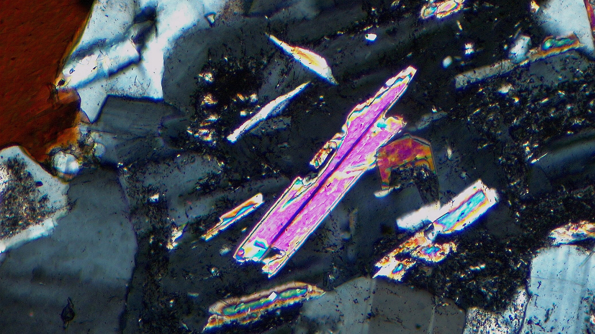

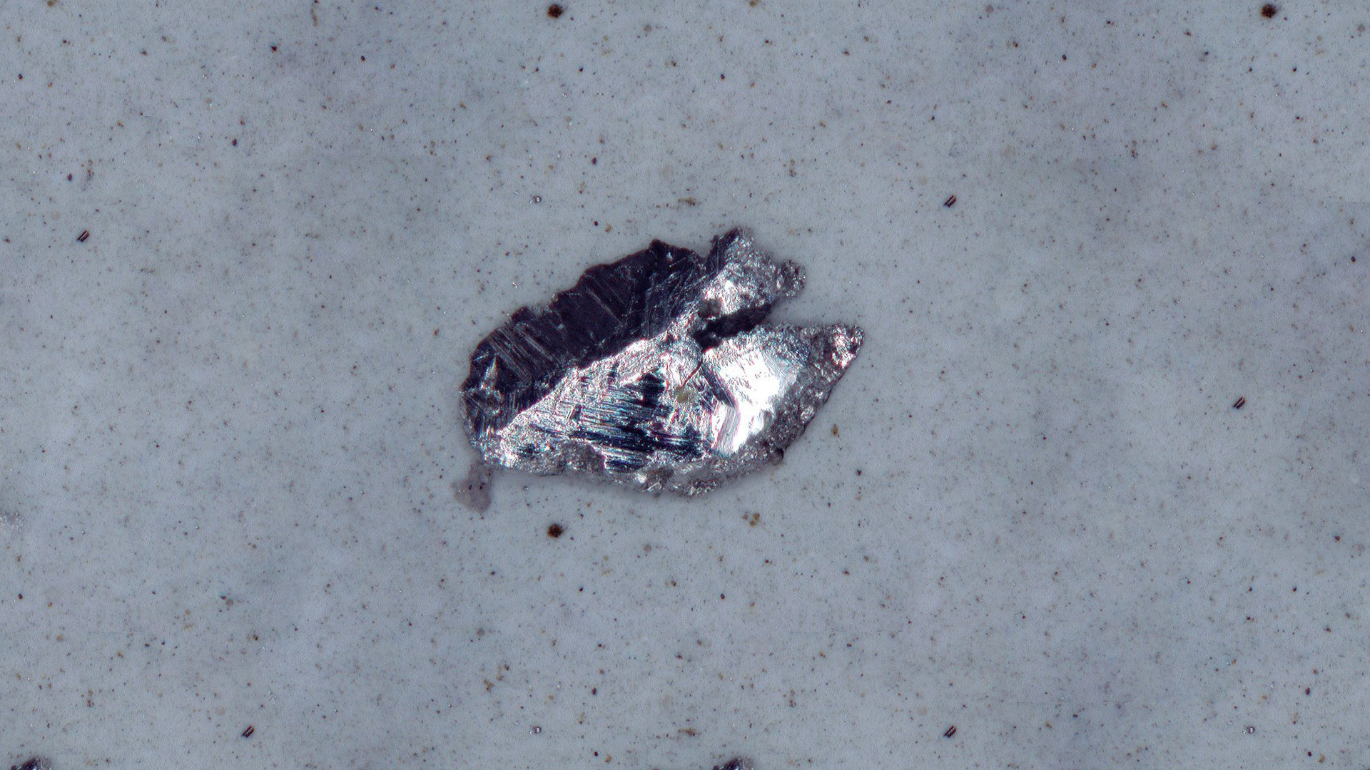
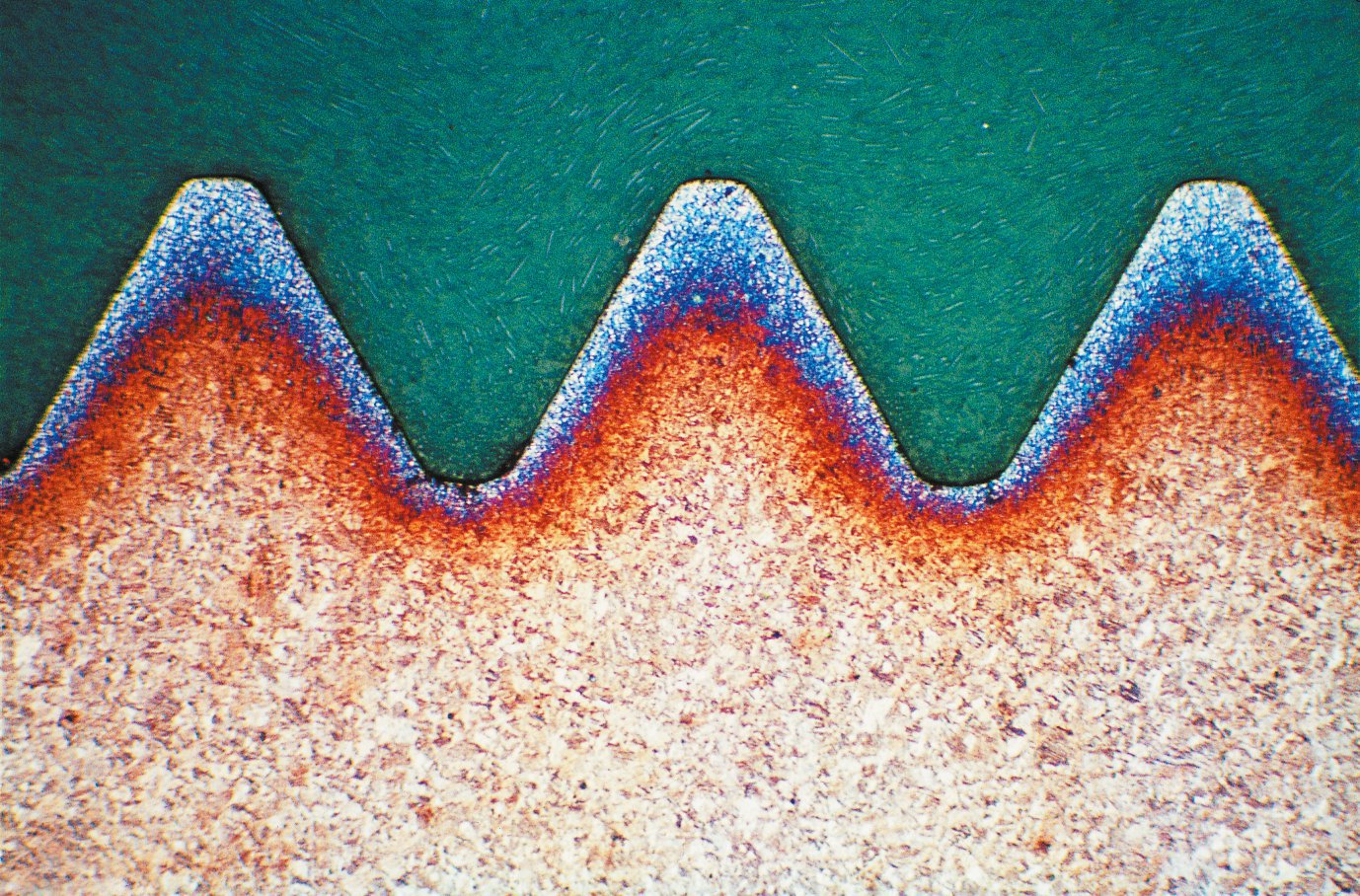
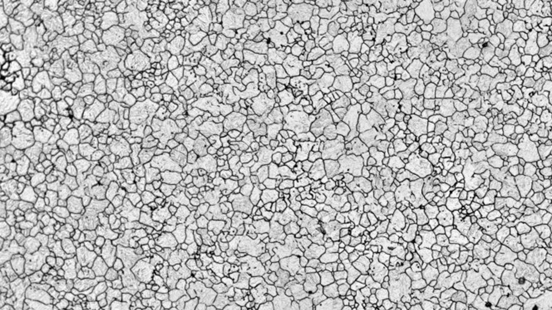
不純物や内部構造の分析方法は?
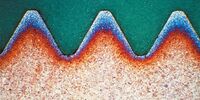
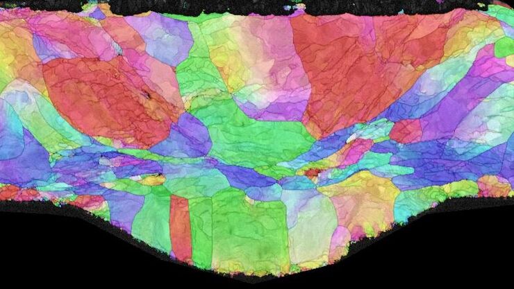
偏光と微分干渉コントラスト(DIC)を備えた顕微鏡を使用することで、標準的な明視野照明では見えないマイクロクラックや介在物のような材料内部の構造を可視化することができます。偏光は異方性物質を強調し、DICは染色することなく透明な試料のコントラストを強調します。DM4 PとDM6 Mの顕微鏡は偏光とDICが可能です。

顕微鏡用の試料はどのようにして調整準備しますか?
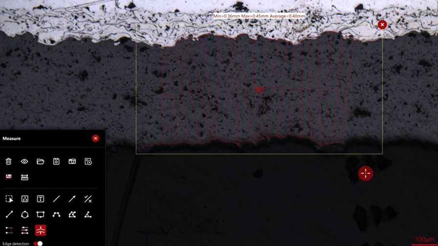
硬質材料の精密な切断、研削、研磨により、ミクロンレベルの精度で特定のサンプル領域をターゲットにすることができます。EM TXPターゲット断面試料作製システムを使用すれば可能です。
EM TIC 3Xは、トリプルイオンビームミリングを採用し、硬質、軟質、脆性、不均質、感熱性など、さまざまなタイプの試料を高品質で表面加工します。

マテリアルサイエンス用顕微鏡の用途は?
マテリアルサイエンス用顕微鏡は、品質管理(QC)、研究開発(R&D)、清浄度・コンタミ解析、故障解析(FA)など、さまざまな用途に使用されます。自動車、航空宇宙、合金、半導体、電子機器、医療機器、地球科学、科学捜査、化学工学、薬学など、さまざまな産業や分野で重要な役割を担っています。

2-Methods-in-1ソリューションが選ばれる理由とは?
2つの手法を1台に統合したソリューションで、光学顕微鏡として使用できるだけでなく、レーザー誘起ブレークダウン分光法(LIBS)による分析も可能です。試料を画像化し、相や介在物のような微細構造を含む化学組成を分析することができます。このソリューションは効率的な材料分析を実現するのに役立ちます。
ライカの顕微鏡がマテリアルサイエンス& 分析に使用される理由とは?

マテリアルサイエンスへのソリューション
DM 750 P | Visoria M& P | DM4 M& P | DM6 M LIBS | DMi8 A | |
| 対物レボルバ | 手動 | コーディング | コーディング | 電動式 | コーディング |
| コントラスト法 | 手動 | コーディング | 電動式 | 電動式 | 電動式 |
| 照明制御 | なし | 利用可能 | 利用可能 | 利用可能 | 利用可能 |
| 観察方法 | IL:BF, DF, Pol, (蛍光) TL:BF、DF、位相差、Pol | IL:BF、DF、Pol、DIC、斜照明(蛍光) TL:BF、DF、位相差、DIC、Pol | IL:BF、DF、Pol、DIC、(蛍光) TL:BF、DF、位相差、DIC、Pol | IL:BF、DF、Pol、DIC、(蛍光) TL:BF、DF、位相差、DIC、Pol | IL:BF、DF、Pol、DIC、(蛍光) TL:BF、DF、位相差、DIC、Pol |
| Zドライブ | 手動 | 手動 | マニュアル/電動式 | 電動式 | 電動式 |
| 回転ステージ | 手動 | 手動 | 手動 | 手動 | 電動式 |
| 対応ソフトウェア | LAS X Industry | Enersight / LAS X インダストリー* | LAS X Industry | LAS X Industry | LAS X Industry |
* LAS X Industry は、Visoria M& P の顕微鏡接続の一部機能を制限しています
関連記事

Visualizing Photoresist Residue and Organic Contamination on Wafers

Rapidly Visualizing Magnetic Domains in Steel with Kerr Microscopy

Quality Assurance Improvement Across Industries
-b-poly(isoprene). Right: Poly(styrene)-b-poly(methyl methacrylate).")
Ultramicrotome Sectioning of Polymers for TEM Analysis

Polarizing Microscope Image Gallery

Revealing Sodium Battery Degradation via Cryo-EM and CryoFIB

A Guide to Polarized Light Microscopy
Workflow Solutions for Sample Preparation Methods for Material Science

Battery Particle Detection During the Production Process

Key Factors for Efficient Cleanliness Analysis

Quality Control via Cross Sections of PCBs, PCBAs, ICs, and Batteries

Five Inverted-Microscope Advantages for Industrial Applications
 stained with osmium tetroxide (OsO4), sectioned with a DIATOME diamond knife at room temperature, and then imaged with HAADF TEM.")
Ultramicrotomy Techniques for Materials Sectioning
 chip cross section acquired at higher magnification showing a region of interest.")
Structural and Chemical Analysis of IC-Chip Cross Sections

How to Prepare and Analyse Battery Samples with Electron Microscopy

Alternative Fuels and Why Sustainable Solutions are Important

Technical Cleanliness in the Automotive Industry for Electromobility

3 Factors Determine the Damage Potential of Particles
マテリアルサイエンス& 分析に関するよくある質問
材料科学は、金属、セラミックス、ポリマー、複合材料を含む材料の構造、特性、性能、加工を研究する学問です。材料の組成や構造が、さまざまな環境下での挙動にどのような影響を与えるかを理解することに重点を置いています。
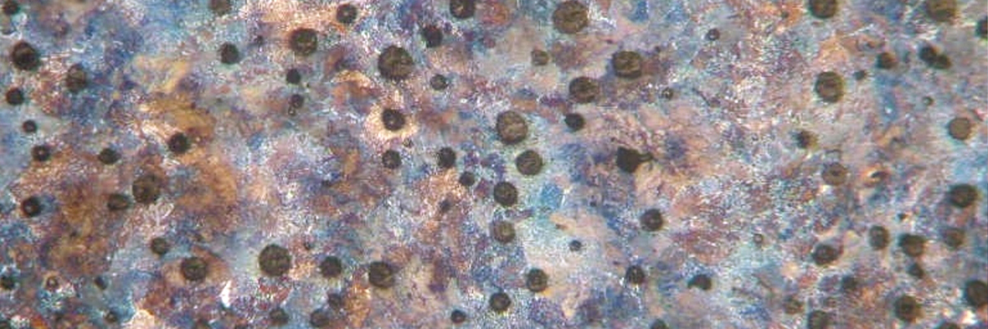
顕微LIBS元素分析システムは、光学とレーザー誘起ブレークダウン分光法(LIBS)を組み合わせ、材料の目視検査と化学組成分析を同時に行います。
LIBSは元素組成の分析手法の一つです。LIBSの原理は以下です。高エネルギーのレーザーパルスを分析対対象物表面に照射します。レーザーエネルギーは吸収され、アブレーションが起こり、クレーターが形成されます。電子励起状態になった原子やイオンを含むプラズマが発生します。これらの原子が基底状態に戻るときに、元素特有の光の波長を放出します。元素が高温励起により発生する光について固有の周波数を有するため、測定波長範囲と感度のみに依存して元素の測定が可能となります。
マテリアル分析は、材料の物理的および化学的特性を測定するために行われます。例えば、鉄鋼やアルミニウムなどの金属合金、ガラスやシリコンなどのセラミックス、プラスチックやポリマー、鉱物や地質サンプルなどです。材料科学や地球科学、品質管理、生産、故障解析、研究開発などによく使用されます。