


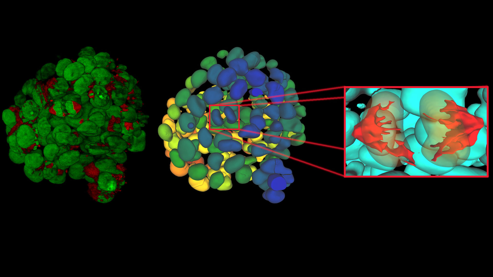

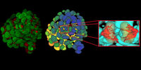


 and co-stained for nuclear DNA (Hoechst 33342), microtubules (Alexa 555) and F-actin (ATTO 643). Image was captured on Mateo FL.")
.")
.")
新しいFlexacam C3顕微鏡カメラ
最新の記事を読む
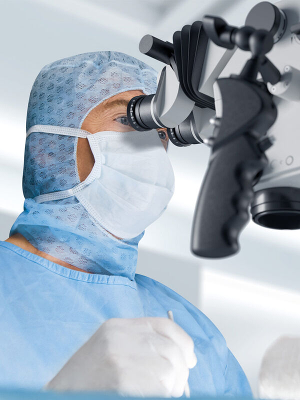
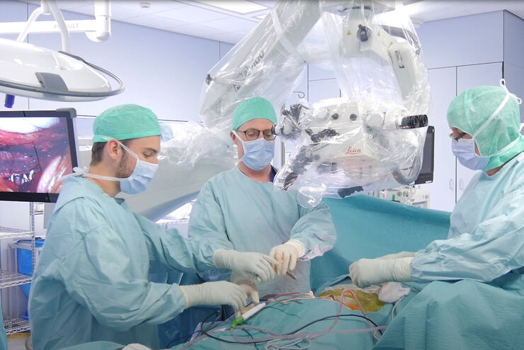
Advanced Visualization in Head and Neck Reconstructive Surgery
PRS surgeons and maxillofacial specialists face unique challenges in reconstructive procedures, particularly when operating in deep, narrow cavities and working with delicate tissues. Achieving…
Advanced Visualization: Transforming Minimally Invasive Spine Surgery
Over the past decade, spine surgery has evolved rapidly with the adoption of minimally invasive surgery (MISS) techniques. These approaches reduce surgical trauma, speed up recovery, and help improve…
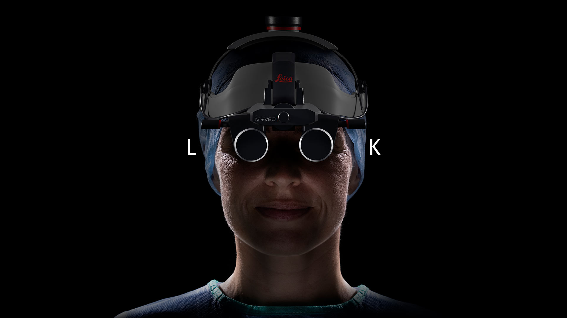
Advanced Visualization in ENT Surgery: Prof. Darrouzet’s Expert Insights
Otologic surgery demands exceptional precision when working within the confined spaces of the middle and inner ear. Procedures such as tympanoplasty, mastoidectomy, and cochlear implant placement…
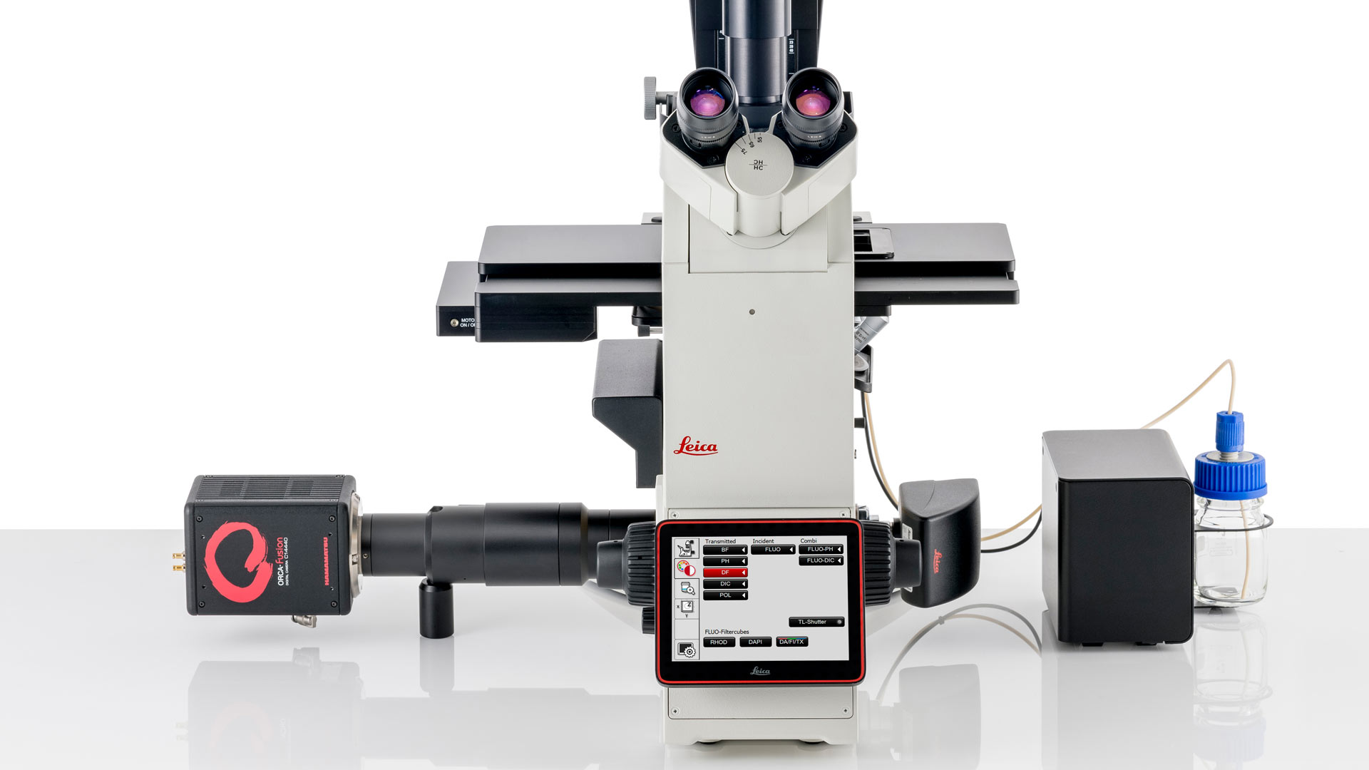
Infinity Optical Systems - From “Infinity Optics” to the Infinity Port
“Infinity Optics” is the concept of a light path with parallel rays between the objective and tube lens of a microscope [1]. Placing flat optical components into this “infinity space” which do not…
Six Features to Consider when Choosing a Dental Microscope
The dental surgical microscope has become increasingly important for high-quality and successful dental medicine, particularly in the field of endodontics. A dentist can conduct micro-invasive…
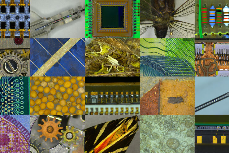
Quality Assurance Improvement Across Industries
Precision is paramount. Imagine a pacemaker that fails mid-operation or a semiconductor flaw that causes a critical system crash. In industries, such as medical devices, electronics, and…
AI meets Deep Visual Proteomics (DVP) to Advance Disease Research
In this webinar, Dr. Andreas Mund will introduce a cutting-edge platform that merges Deep Visual Proteomics (DVP) with AI-powered pathology models, enabling high-resolution mapping of key regions in…
Microscopy and AI Solutions for 2D Cell Culture
This eBook explores the integration of microscopy and AI technologies in 2D cell culture workflows. It highlights how traditional imaging methods—such as brightfield, phase contrast, and…
AI-Powered Hi-Plex Spatial Analysis Tools for Breast Cancer Research
Breast cancer (BC) is the leading cause of cancer-related deaths in women. Investigating the tumor microenvironment (TME) is crucial to elucidate the mechanisms of tumor progression. Systematic…
Focus on Long-Term Imaging in 3D with Light Sheet Microscopy
Long-term 3D imaging reveals how complex multicellular systems grow and develop and how cells move and interact over time, unlocking critical insights into development, disease, and regeneration.…
