Aivia AI Image Analysis Software
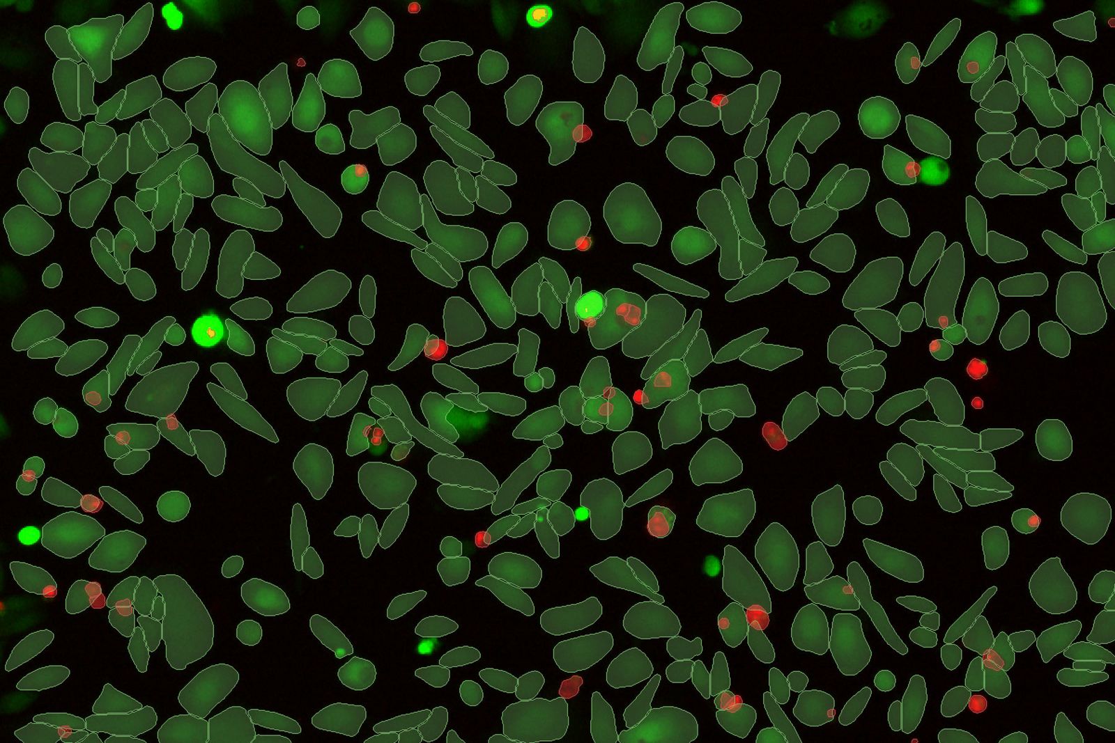
A complete analysis workflow from accurate deep-learning based cell segmentation to automatic phenotyping and data exploration for 3D multiplexed images.
Subjectivity of analysis and poor reproducibility are key hurdles to be overcome for biological image analysis. Standard segmentation can lead to sub-standard results and require substantial manual curation which is subject to human error. Aivia changes all of that.
マルチウェルデータを可視化するインタラクティブチャート
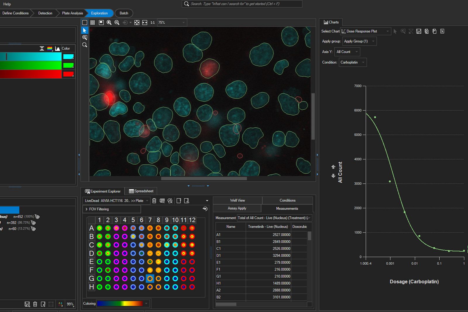
マルチウェル実験データを高度に探索・解析Aivia 16は、集約サマリーチャートと統計解析向けデータエクスポート機能を備え、マルチウェル実験データの探索を強化。より深いインサイトの発見を支援します。
- ウェル別、オブジェクトセット別、条件別に集約したマルチウェルデータから、価値あるインサイトを取得
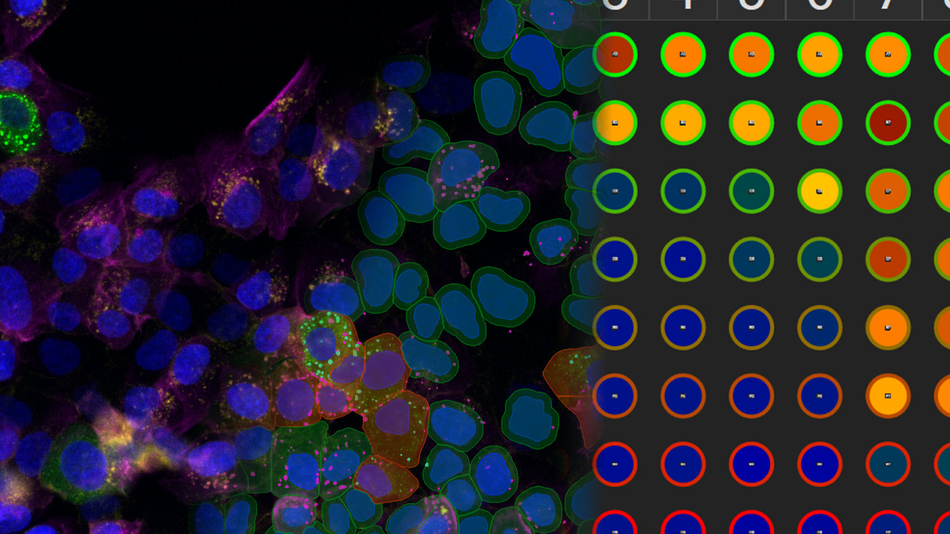
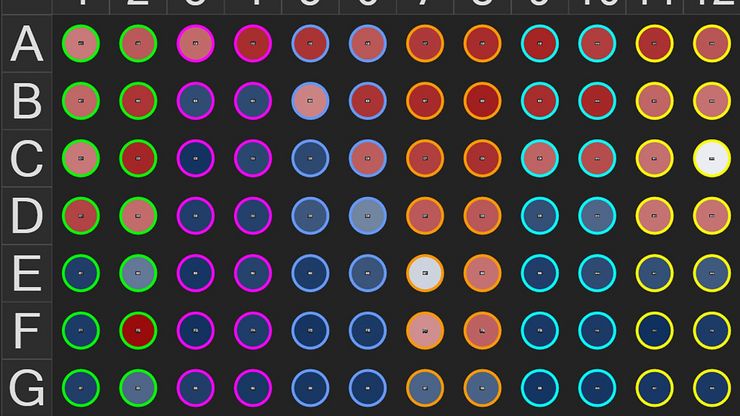
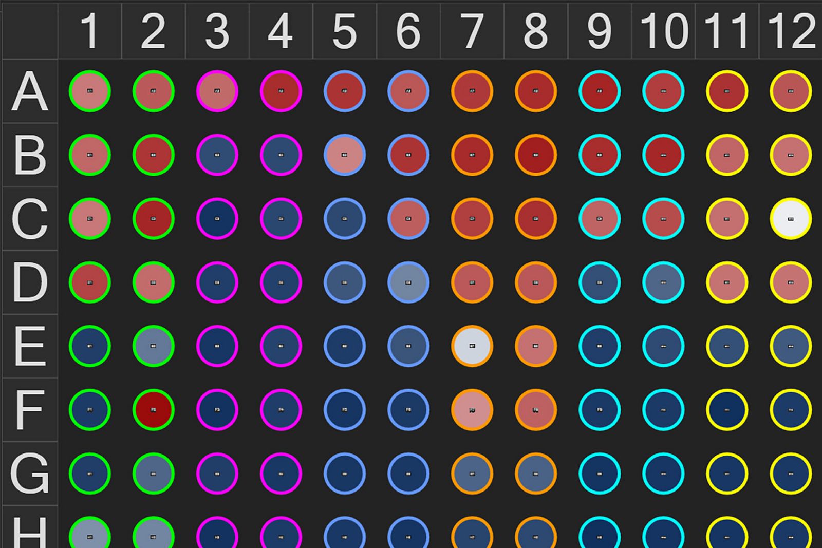
- Experiment Explorerのマルチウェルヒートマップにより、ウェルごとの解析結果を一覧表示し、目的のデータへ迅速にアクセスできます。
- Summary Chartsでデータの集約・表示方法を柔軟にカスタマイズ
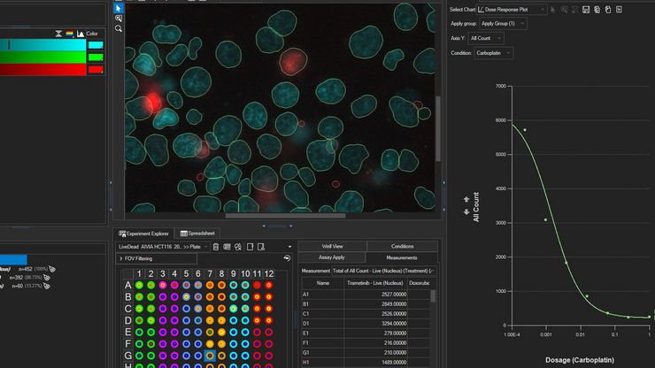
- 用量反応曲線とIC50/EC50自動算出により、マルチウェル実験における細胞応答を明らかにします。
- 統合されたデータを統計解析ソフトウェアへエクスポートすることで、より高度なデータ解析を実行できます。
AI access for all
Aivia makes advanced data analysis accessible for all biologists - with no computer science expertise required.
The Aivia platform has been designed with the end-user in mind. This means with Aivia you can quickly and reliably generate high-quality results. The Aivia platform includes all state-of-the-art applications you will need in a unified user experience.
Quickly train laboratory users on the platform, to conduct their analysis without any specialist expertise.
- Speed up your imaging projects and publish faster
- Benefit from next-generation, easy to use machine learning segmentation and classification tools
- Conduct parameter-free image segmentation
- Easily train, update and apply deep learning models using local resources or the AiviaCloud platform
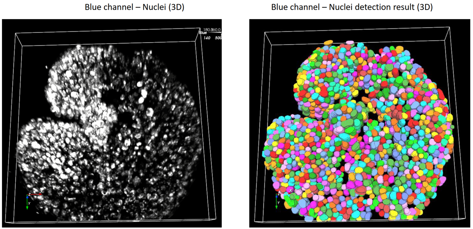
Total freedom on a single platform
Aivia's powerful and fast 2-5D visualization and analysis unlocks all the value of your data - within a single platform.
No longer does your team have to learn to operate and adopt multiple imaging and analyses systems into their workflow - the Aivia platform unifies all state-of-the-art applications you will need in a unified user experience. Aivia can leverage both local and cloud computing resources. You can install and use Aivia both on your local computer as well as via a web browser, AiviaWeb. Aivia works seamlessly with all microscopy imaging systems.
Your team can also access all files created by your imaging systems anywhere - all you need is an internet connection.
- Powerful and fast 2-5D visualization and analysis - accessible anywhere
- Includes 22 applications and 20 pre-trained deep learning models (image segmentation, restoration and virtual staining)
- Reliable and easy to use cloud access with flexible IT architectures supported
- Over 45 microscopy file formats supported

Start a free trial
Using state-of-the-art, AI-first software architecture, Aivia is a uniquely innovative and complete 2-to-5D image visualization, analysis and interpretation platform designed for the reliable processing and reconstruction of highly complex images in just minutes.
- Make AI-enhanced image analysis accessible for all - with no computer science expertise required
- Leverage machine learning capabilities to generate robust and reproducible segmentation results
- Realize powerful and fast 2-5D visualization and analysis to unleash the value of your data - all within a single platform
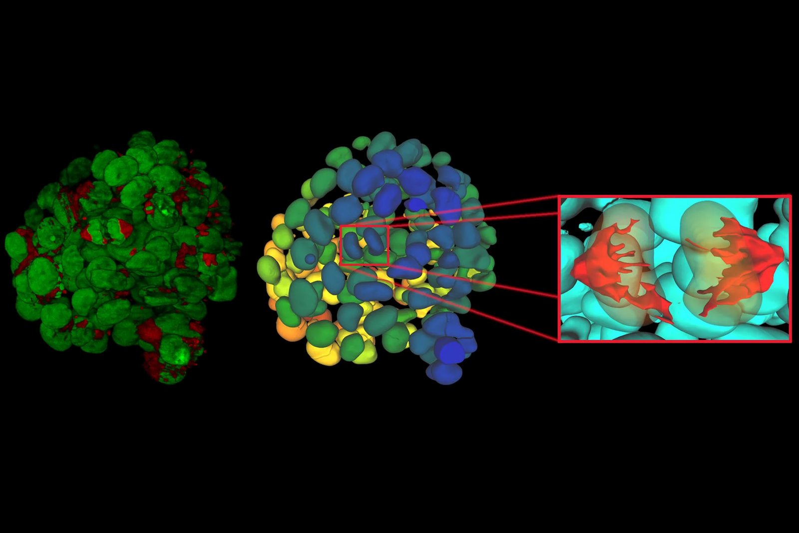
Explore Aivia subscription plans
Aivia is available via subscription, giving you the flexibility to select a plan that fits your lab’s needs now, or in the future as your research needs evolve.
With technical support and free software upgrades for the duration of your subscription, you will always have the lasted AI-powered analysis tool for your research.
- Go - The essential tools to get you started with AI image analysis
- Elevate - Take your AI image analysis to the next level with CellBio or Neuro
- Apex - A comprehensive AI image analysis platform for labs and core facilities
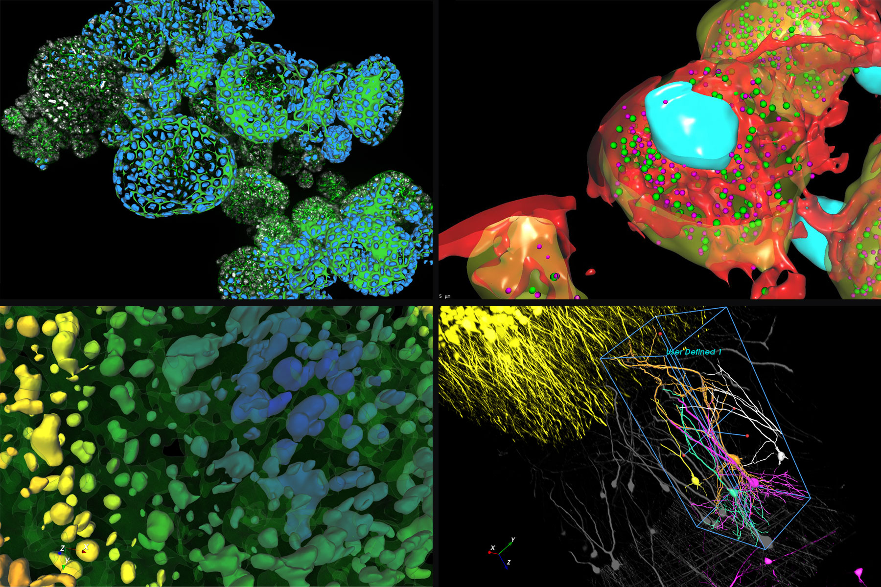
Everything you need to start analyzing images
Aivia Go offers state-of-the-art image visualization and analysis tools in a single platform, including multiple AI-powered features, to meet your challenging image visualization and analysis needs. Simple segmentation workflows and batch processing capabilities yield results faster, helping you leap from data to publication.
Aivia Go supports a large range of applications from 2D to 5D image analysis tasks such as detection and tracking. It is the ideal package for research laboratories and imaging core facilities. No computer expertise required.
- AI-powered classifiers
- 12+ image analysis recipes
- 2D -5D image visualization
Features
Image Analysis Recipes - Deploy a wide range of 2D to 5D image analysis recipes for the most popular analysis applications:
- Count cells, nuclei and particles in 2D and 3D
- Track cells (in phase contrast and fluorescence), nuclei, and particles in 2D and 3D
- Cell proliferation and wound healing assays (phase contrast)
- Neurite outgrowth
- Stem cell colony detection (phase contrast)
- Cell tracking (phase contrast)
Descubrimiento rápido de información en experimentos multipocillos complejos
Aivia Accelerate permite una rápida exploración de las relaciones entre variables clave en experimentos multipocillos complejos, permitiendo a los científicos extraer información procesable de los datos de imagen con una configuración mínima y sin necesidad de codificación.
Características
- Analice los resultados agregados en todas las condiciones experimentales utilizando gráficos de resumen con estadísticas de población
- Acelere los análisis con flujos de trabajo preconfigurados
- Despliegue de un algoritmo generalista de segmentación de células aprendizaje profundo preentrenado para la segmentación por ejemplos
- No se requiere entrenamiento de modelos, codificación ni experiencia en IA
- Clasifique automáticamente las poblaciones celulares
- Generación automática de informes exhaustivos
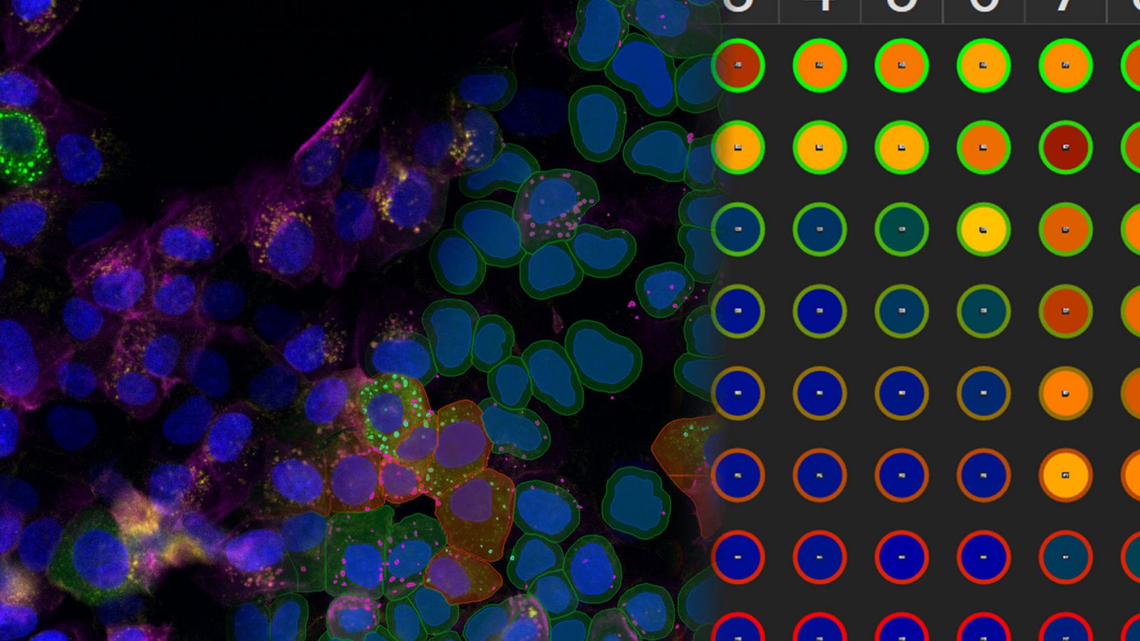
Take your AI image analysis to the next level with Aivia’s simple workflows
A complete solution for research laboratories and core imaging facilities, Aivia Elevate subscribers can choose specialized AI solutions for either neuro or cell biology image visualization and analysis.
Analyze cellular interactions and measure the distances between different compartments down to the channel level with Elevate for CellBio . It includes tools to perform accurate cell detection in 2D multiplexed images, identify cell phenotypes and explore spatial insights. Or automate challenging neuroimaging tasks, such as neuron tracing, dendrite segmentation, and more with Elevate for Neuro.
Want both options? Upgrade to Aivia Apex for full functionality plus the ability to apply your own deep learning models.
Features
Cell or Neuron Analysis Recipes - Choose between specialized analysis tools for neuroscience or cell biology research
- Neuron Analysis Recipes (Fluorescence/EM): Detect and trace neurons in 3D data sets
- Cell Analysis Recipes (2D/3D): Observe complex cellular interactions in 2D and 3D data sets
A comprehensive AI image analysis platform for labs and core facilities
Aivia Apex is a comprehensive microscopy image analysis solution for researchers who need a variety of image analysis applications. Apex also provides microscopists with the flexibility to apply their own deep learning models, third party models or open-source repositories.
Ideal for large research groups and core imaging facilities, Aivia Apex’s Floating License Manager allows a site to run all licenses on any machine on their local network.
- Everything in Elevate Neuro & CellBio
- Expanded deep learning capabilities
- Floating license manager
Features
Image Analysis Recipes - Deploy a wide range of 2D to 5D image analysis recipes for the most popular analysis applications
- Nuclei count and tracking
- Cell count and tracking
- Particle count and tracking
- Cell proliferation assay
- Neurite outgrowth
- Wound healing (phase contrast)
- Stem cell colony detection (phase contrast)
- Cell tracking (phase contrast)
- 3D Object Analysis and Tracking recipes including neuron and cell analysis
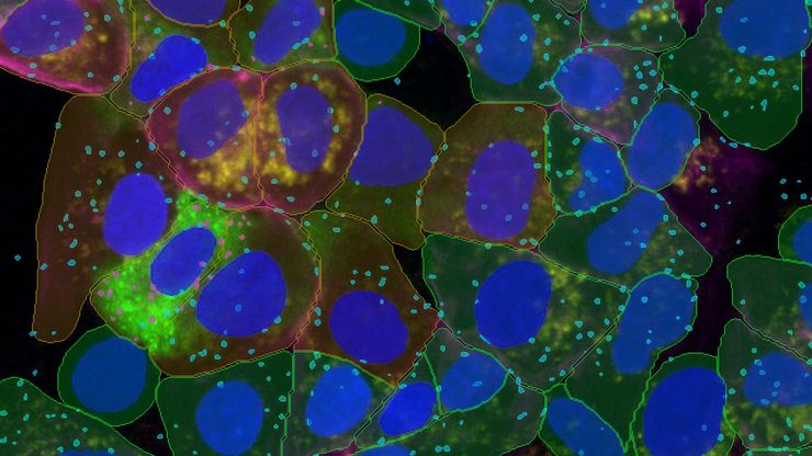
進化したSegment by Example
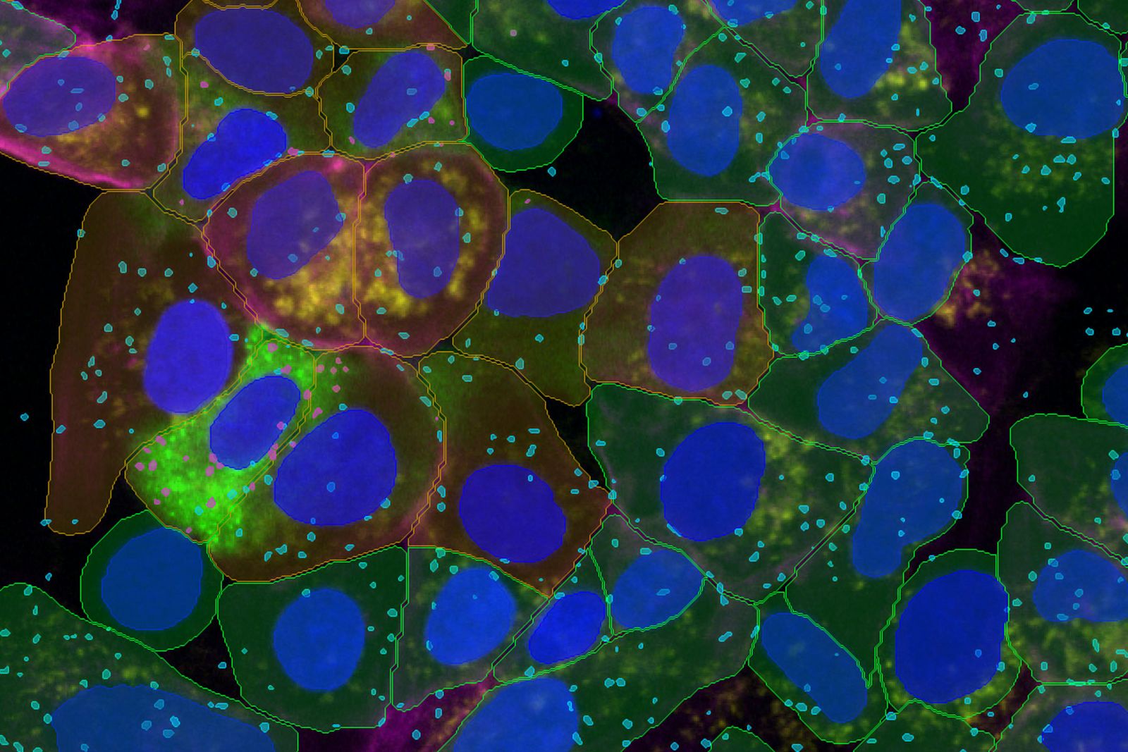
最小限の入力で、より深いインサイトをより速く。Aivia 16の「Segment by Example」は、特許出願中のAivia独自ディープラーニングアルゴリズムを採用。わずかな操作で高精度な細胞検出を可能にし、使いやすさと解析精度を新たなレベルへ引き上げます。
- たった3つのサンプルで学習し、2D・3D画像の細胞を高精度にセグメント化。
- 複数の画像チャネルを組み合わせた2D/3Dマルチプレックス細胞検出
- 2D画像内の細胞、核、および複数種類の小胞を自動セグメンテーション
- 異なる細胞集団のフェノタイプを一度に解析
- 細胞および核の検出速度を最大12.8倍向上
マルチウェルデータを簡単にインポート
マルチウェルデータを簡単にインポート。Aivia 16により、研究者はデータを簡単に取り込み、直感的に探索し、高度な解析をシームレスに実行できます。Mica、DMi8、STELLARISで取得したLIF/XELFファイルから、マルチウェル実験データを直接取り込み、すぐに解析を開始できます。。
新しくなったExperiment Explorerを使用して、マルチウェル実験のウェルや視野をシームレスにナビゲートし、Aiviaに読み込まれたデータを迅速にレビューできます。
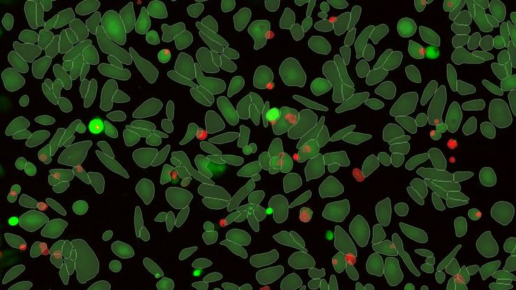
ガイド付きアッセイ解析
アッセイ解析をEnd-to-Endでシンプルにガイド付きアッセイ解析ワークフローにより、データ取得からインサイトの発見までをシンプルかつ効率的に実現。マルチウェルサンプルにおいて優れた精度と再現性を提供し、データの価値を最大限に引き出すためのクラス最高レベルの解析性能を実現します。
- Analysis Launchpadからガイド付きアッセイ解析ワークフローを起動
- Guided Sequenceにより、実験条件の設定から細胞検出、フェノタイピング、バッチ解析までをシームレスに実行できます。
- 細胞数カウント、形態解析、Live/Deadアッセイ、アポトーシス解析など、代表的なアッセイ向けに最適化された4種類のGuided Sequenceを利用できます。
- ウェル別、条件別、プレート別に解析結果を集約・比較
- サンプル画像を用いて解析パイプラインを調整し、実験全体へシームレスに反映