Industriel
Industriel
Plongez dans des articles détaillés et des webinaires consacrés à l'inspection efficace, à l'optimisation des flux de travail et au confort ergonomique dans les contextes industriels et pathologiques. Les sujets abordés comprennent le contrôle de la qualité, l'analyse des matériaux, la microscopie en pathologie, parmi beaucoup d'autres. C'est ici que vous obtiendrez des informations précieuses sur l'utilisation des technologies de pointe pour améliorer la précision et l'efficacité des processus de fabrication, ainsi que pour établir des diagnostics et des recherches pathologiques précis.
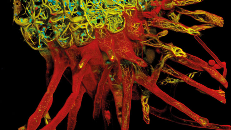
Benefits of TauContrast to Image Complex Samples
In this interview, Dr. Timo Zimmermann talks about his experience with the application of TauSense tools and their potential for the investigation of demanding samples such as thick samples or…
Fast, High-quality Vitrification with the EM ICE High Pressure Freezer
The EM ICE High Pressure Freezer was developed with a unique freezing principle and uses only a single pressurization and cooling liquid: liquified nitrogen (LN2). This design enables three major…
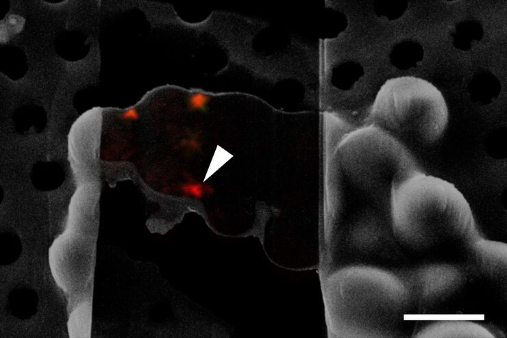
Targeting Active Recycling Nuclear Pore Complexes using Cryo Confocal Microscopy
In this article, how cryo light microscopy and, in particular cryo confocal microscopy, is used to improve the reliability of cryo EM workflows is described. The quality of the EM grids and samples is…
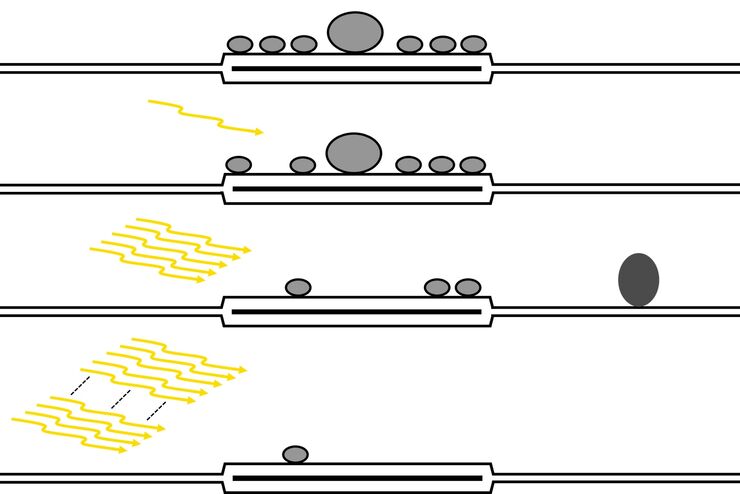
Investigating Synapses in Brain Slices with Enhanced Functional Electron Microscopy
A fundamental question of neuroscience is: what is the relationship between structural and functional properties of synapses? Over the last few decades, electrophysiology has shed light on synaptic…
The Power of Pairing Adaptive Deconvolution with Computational Clearing
Learn how deconvolution allows you to overcome losses in image resolution and contrast in widefield fluorescence microscopy due to the wave nature of light and the diffraction of light by optical…
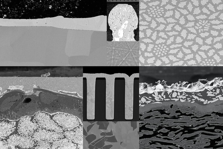
Ion Beam Milling Guide: Enhancing Surface Quality for High-Resolution Imaging and Analysis
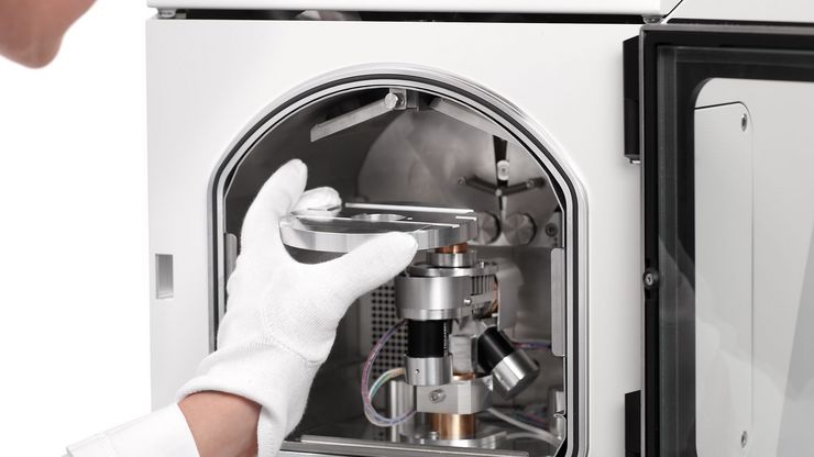
In this article you can learn how to optimize the preparation quality of your samples by using the ion beam etching method with the EM TIC 3X ion beam milling machine. A short introduction of the…
Solutions de pulvérisation cathodique et de cryofracture
Du dépôt en vide secondaire à température ambiante à la pulvérisation cryogénique sous vide poussé, Leica Microsystems couvre la gamme complète des besoins en matière de pulvérisation.
Studying Pulmonary Fibrosis
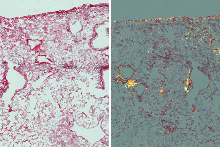
The results shown in this article demonstrate that fibrotic and non-fibrotic regions of collagen present in mouse lung tissue can be distinguished better with polarized light compared to brightfield.…
Image Gallery: THUNDER Imager
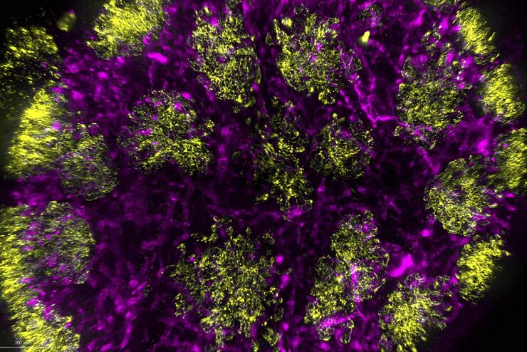
To help you answer important scientific questions, THUNDER Imagers eliminate the out-of-focus blur that clouds the view of thick samples when using camera-based fluorescence microscopes. They achieve…
