STELLARIS DIVE
共焦点顕微鏡
製品紹介
Home
Leica Microsystems
STELLARIS DIVE 多光子顕微鏡
マルチカラー多光子顕微鏡でより真実に近く
最新の記事を読む

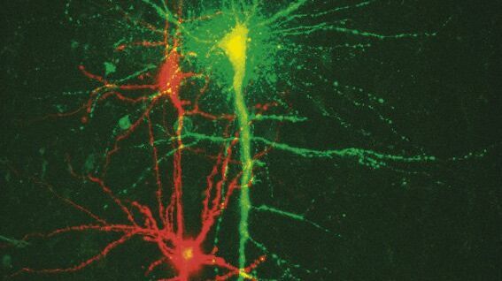
Multicolor Image Gallery
Fluorescence multicolor microscopy, which is one aspect of multiplex imaging, allows for the observation and analysis of multiple elements within the same sample – each tagged with a different…

How to Quantify Changes in the Metabolic Status of Single Cells
Metabolic imaging based on fluorescence lifetime provides insights into the metabolic dynamics of cells, but its use has been limited as expertise in advanced microscopy techniques was needed.
Now,…

研究におけるモデル生物
モデル生物とは、特定の生物学的プロセスを研究するために研究者が使用する生物種です。 モデル生物は、人間と似た遺伝的特徴を持ち、遺伝子学、発生生物学、神経科学などの研究分野で一般的に使用されています。 通常、モデル生物は実験環境での維持や繁殖が容易であること、生殖サイクルが短いこと、または、特定の形質や病気を研究するために突然変異体を生成する能力を持つことで選ばれます。

LIGHTNINGによって試料から最大限の情報を引き出す
LIGHTNINGは、他の方法では簡単に可視化できない、微細な構造や形態を完全自動で明らかにする、適応能力に優れた情報抽出プロセスです。 LIGHTNINGは、画像全体を同一のパラメーターで演算する従来型の手法とは異なり、ボクセル(3次元画素)ごとに適切なパラメーターを算出することによって、最高の忠実度であらゆる微細形態を明らかにします。

ウイルス学
ウイルス研究のためのイメージングと試料作製ソリューション
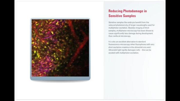
Principles of Multiphoton Microscopy for Deep Tissue Imaging
This tutorial explains the principles of multiphoton microscopy for deep tissue imaging. Multiphoton microscopy uses excitation wavelengths in the infrared taking advantage of the reduced scattering…
New Standard in Electrophysiology and Deep Tissue Imaging
The function of nerve and muscle cells relies on ionic currents flowing through ion channels. These ion channels play a major role in cell physiology. One way to investigate ion channels is to use…

高度な組織イメージングおよび解析
Leica Microsystems の高度なイメージングソリューションを活用することで、組織の構造および機能に対する理解を深め、空間生物学や疾患機構の解明を促進します。
Fields of Application
高度な組織イメージングおよび解析
Leica Microsystems の高度なイメージングソリューションを活用することで、組織の構造および機能に対する理解を深め、空間生物学や疾患機構の解明を促進します。
