is mobile? false
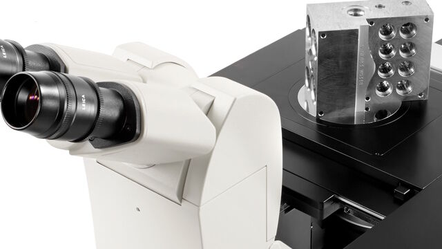
電顕用試料作製装置
電顕用試料作製装置
ライカマイクロシステムズの電顕用試料作製装置は、医学・生物学用、産業用共に幅広い製品群で、様々なニーズにお応えします。
Leica Science Lab Show subnavigation
最新の記事を読む
ライカマイクロシステムズのサイエンスラボポータル は、顕微鏡をテーマとする科学研究や記事を提供しています。 コンテンツは、日常業務や実験で、ビギナーから経験豊富な専門家、科学者まで幅広くサポートします。
電顕用試料作製装置
ライカマイクロシステムズの電顕用試料作製装置は、医学・生物学用、産業用共に幅広い製品群で、様々なニーズにお応えします。
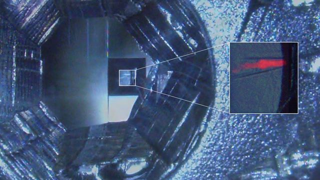
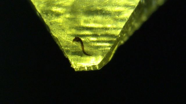
How Fluorescence Guides Sectioning of Resin-embedded EM Samples
Electron microscopes, including transmission electron microscopes (TEM) and scanning electron microscopes (SEM), are widely utilized to gain detailed structural information about biological samples or non-living materials. Ultramicrotomy is the preferred technique for producing ultrathin sections, less than 100 nm thick for TEM/SEM analysis. During sample preparation small sample pieces are embedded in epoxy or acrylic resin, excess resin is trimmed away, and the specimen is sliced into ultrathin sections (50 nm - 100 nm) using a glass or diamond knife.
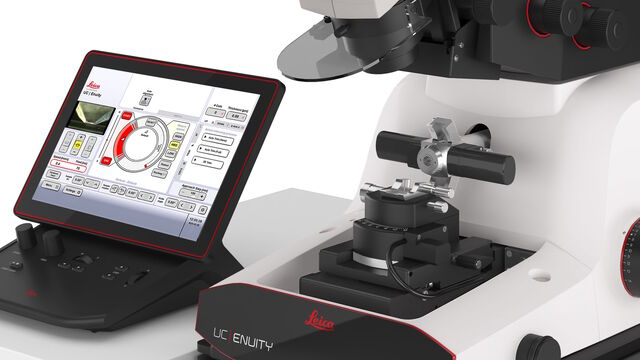
How to Save Time and Samples by Automated Ultramicrotomy
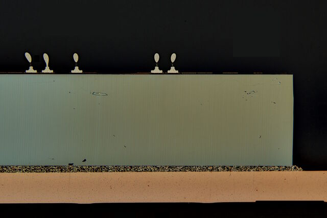
This article describes how 3D micro-CT data of a resin-embedded electron microscopy sample can be used to trim the specimen down to a defined target plane prior to sectioning. The interactive and automated approach described using Leica Microsystems’ UC Enuity saves time, reduces sample loss and training time for unexperienced users.
From Bench to Beam: A Complete Correlative Cryo Light Microscopy Workflow
In the webinar entitled "A Multimodal Vitreous Crusade, a Cryo Correlative Workflow from Bench to Beam" a team of experts discusses the exciting world of correlative workflows for structural biology that empower researchers to study fine details of biological structures. Watch and explore the latest developments, instruments, and techniques in cryo workflows for correlative light electron microscopy (cryo-CLEM).

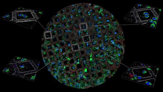
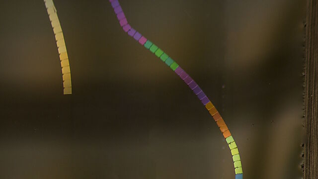
How to Automatically Obtain Fluorescent Cells of Interest in a Block-face
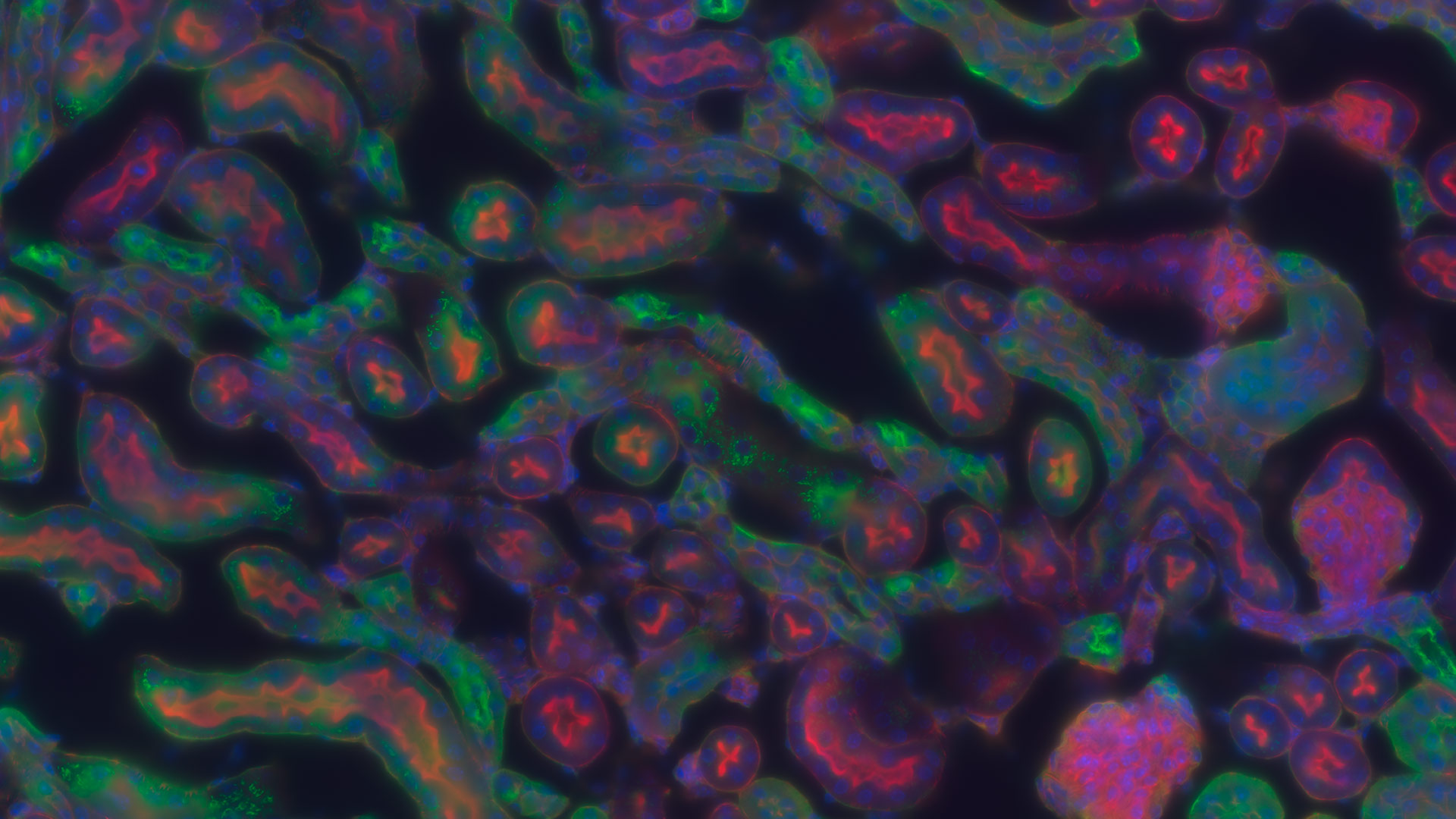
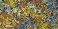
Block-face created by automatic trimming under fluorescence.
Mammalian cells of interest, stained with CellTrackerTM Green are visualized within the block-face using the UC Enuity equipped with the stereo microscope M205 FA. In the background a carbon finder grid in black is visible.
Improve Your Ultramicrotomy Workflow with Automated Sectioning
Discover advanced digital ultramicrotomy tools for fast and accurate automated sectioning. Learn about autoalignment, and efficient sample trimming leveraging 3D µCT data. See application examples that advance EM sample preparation workflows. Watch now to enhance your lab's precision and efficiency.
Workflow Solutions for Sample Preparation Methods for Material Science
This brochure presents and explains appropriate workflow solutions for the most frequently required sample preparation methods for material science samples.
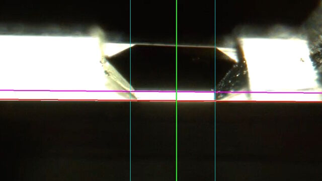
Automatic Alignment of Sample and Knife for High Sectioning Quality
Automatic alignment of sample and knife on the ultramicrotome UC Enuity, enabling even untrained users to create ultrathin sections with reduced risk of losing precious sections.
High Quality Sectioning in Ultramicrotomy
Discover the significance of achieving high-quality uniform sections with ultramicrotomy for precise imaging in electron microscopy.
Rapid Semiconductor Inspection with Microscope Contrast Methods
Semiconductor inspection during the production of patterned wafers and ICs (integrated circuits) is important for identifying and minimizing defects. To increase the efficiency of quality control in the early stages of production and to ensure reliable IC chip performance, microscopy solutions should combine different contrast methods that provide complete and accurate information about different defects. Our free guide details microscopy techniques & optimizing quality control. Get your free copy today!
Five Inverted-Microscope Advantages for Industrial Applications
With inverted microscopes, you look at samples from below since their optics are placed under the sample, with upright microscopes you look at samples from above. Traditionally, inverted microscopes are used for life science research, because gravity makes samples sink to the bottom of a holder with aqueous solution and you don’t see a lot from above.
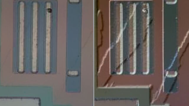
Structural and Chemical Analysis of IC-Chip Cross Sections
This article shows how electronic IC-chip cross sections can be efficiently and reliably prepared and then analyzed, both visually and chemically at the microscale, with the EM TXP and DM6 M LIBS solutions.
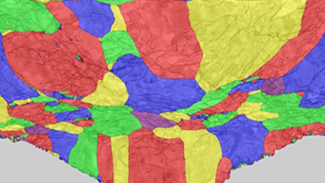
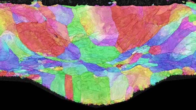
High-Quality EBSD Sample Preparation
This article describes a method for EBSD sample preparation of challenging materials. The high-quality samples required for electron backscatter diffraction are prepared with broad ion-beam milling.
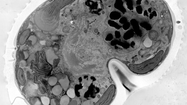
How Marine Microorganism Analysis can be Improved with High-pressure Freezing
In this application example we showcase the use of EM-Sample preparation with high pressure freezing, freeze substiturion and ultramicrotomy for marine biology focusing on ultrastructural analysis of dinoflagellates in particular. The mobility the EM ICE unit allowed high pressure freezing directly at the marine station after sample collection.
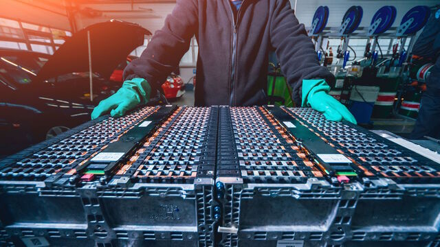
How to Prepare and Analyse Battery Samples with Electron Microscopy
This workshop covers the sample preparation process for lithium and novel battery sample analysis, as well as other semiconductor samples requiring high-resolution cross-section imaging.
New Imaging Tools for Cryo-Light Microscopy
New cryo-light microscopy techniques like LIGHTNING and TauSense fluorescence lifetime-based tools reveal structures for cryo-electron microscopy.
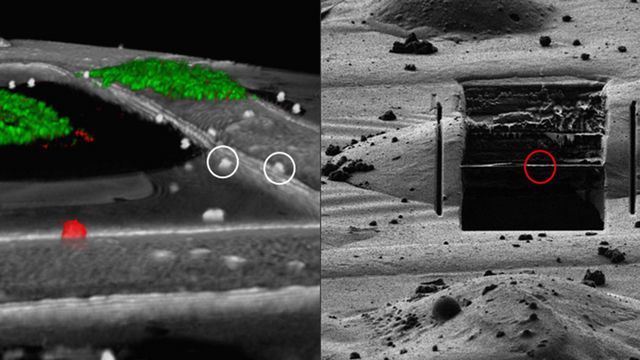
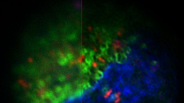
How to Target Fluorescent Structures in 3D for Cryo-FIB Milling
This article describes the major steps of the cryo-electron tomography workflow including super-resolution cryo-confocal microscopy. We describe how subcellular structures can be precisely located in 3D under cryo-fluorescence and how their coordinates are provided for subsequent cryo--FIB milling and cryo-transmission electron microscopy.