Leica EM UC7
Ultramikrotome & Kryo-Ultramikrotome
Elektronenmikroskopie Probenvorbereitung
Produkte
Startseite
Leica Microsystems
Leica 
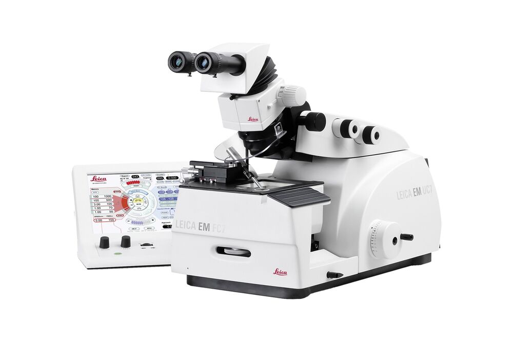
EM UC7 Das Ultramikrotom für ausgezeichnete Schnitte unter Raum- und Tieftemperatur
Lesen Sie unsere neuesten Artikel
How Marine Microorganism Analysis can be Improved with High-pressure Freezing
In this application example we showcase the use of EM-Sample preparation with high pressure freezing, freeze substiturion and ultramicrotomy for marine biology focusing on ultrastructural analysis of…
Exploring the Structure and Life Cycle of Viruses
The SARS-CoV-2 outbreak started in late December 2019 and has since reached a global pandemic, leading to a worldwide battle against COVID-19. The ever-evolving electron microscopy methods offer a…
Investigating Synapses in Brain Slices with Enhanced Functional Electron Microscopy
A fundamental question of neuroscience is: what is the relationship between structural and functional properties of synapses? Over the last few decades, electrophysiology has shed light on synaptic…
Workflows and Instrumentation for Cryo-electron Microscopy
Cryo-electron microscopy is an increasingly popular modality to study the structures of macromolecular complexes and has enabled numerous new insights in cell biology. In recent years, cryo-electron…
Upright Fluorescence Microscopy for Virus Replication Studies
The severe acute respiratory syndrome coronavirus 2 (SARS-CoV-2) emerged in late 2019 and spread over the world quickly. Due to its dramatic impact, researchers are interested in the nature of the…
Microscopy in Virology
The coronavirus SARS-CoV-2, causing the Covid-19 disease effects our world in all aspects. Research to find immunization and treatment methods, in other words to fight this virus, gained highest…
Workflow Solutions for Sample Preparation Methods for Material Science
This brochure presents and explains appropriate workflow solutions for the most frequently required sample preparation methods for material science samples.
Ultrastructural Preservation and Improved Visualization of Membranes in Primary Bovine Chromaffin Cells
Intracellular dynamic events, such as protein trafficking, recycling, and degradation as well as signal transduction, to name a few, can be studied using live-cell imaging techniques. However,…
Introduction to Ultramicrotomy
When studying samples, to visualize their fine structure with nanometer scale resolution, most often electron microscopy is used. There are 2 types: scanning electron microscopy (SEM) which images the…
Superior Ultrastructural Preservation and Structural Contrast in Drosophila Tissue
Optimal structural preservation of tissue can only be obtained by high pressure freezing. However, preparing the samples in optimal conditions is challenging. This article explains in detail how to…
Cryo-Electron Microscopy of Vitreous Sections (CEMOVIS) of Yeast
Application Note for Leica EM HPM100 - The sections are of yeast frozen with a Leica HPM100 high pressure freezer in the copper tube system, the cell paste was mixed with a pH 6.5 MES/dextran buffer…
Perusing Alternatives for Automated Staining of TEM Thin Sections
Contrast in transmission electron microscopy (TEM) is mainly produced by electron scattering at the specimen: Structures that strongly scatter electrons are referred to as electron dense and appear as…
Atomic Force Microscopy Study of a Stretched Impact Copolymer
Atomic force microscopy (AFM) is a powerful tool in the suite of nanoscale characterization techniques that provides a variety of information including topography, mechanical properties, and…
Anwendungsbereiche
Zebrafisch-Forschung
Für optimale Ergebnisse während der Bewertung, Sortierung, Manipulation und Bildgebung von Modellorganismen ist es entscheidend feine Details und Strukturen genauestens zu erkennen. Das bildet die…
Fluoreszenz
Die Fluoreszenz ist eines der am häufigsten verwendeten physikalischen Phänomene in der biologischen und analytischen Mikroskopie, vor allem wegen ihrer hohen Empfindlichkeit und Spezifität. Erfahren…