Kontaktieren Sie uns

Organoide und 3D-Zellkultur

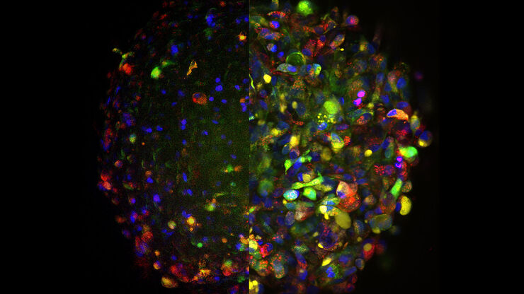
Einer der sensationellesten Fortschritte in der Life-Science-Forschung in jüngster Zeit ist die Entwicklung von 3D-Zellkultursystemen wie Organoiden, Sphäroiden oder Organ-on-a-Chip-Modellen. Eine 3D-Zellkultur ist eine künstliche Umgebung, in der Zellen in allen drei Dimensionen wachsen und mit ihrer Umgebung interagieren können. Diese Bedingungen ähneln denen von In-vivo-Methoden. Organoide sind eine Art 3D-Zellkultur, die organspezifische Zelltypen enthält, die eine räumliche Organisation aufweisen und einige Funktionen des Organs replizieren können.

Organoide bilden weitgehend ein physiologisch relevantes System nach, mit dem Forscher komplexe, mehrdimensionale Fragen wie Krankheitsbeginn, Geweberegeneration und Wechselwirkungen zwischen Organen untersuchen können. Die Lichtmikroskopie ist ein wesentlicher Beitrag zur Untersuchung der mit Organoiden modellierten komplexen Systeme.

Die Bildgebungslösungen von Leica Microsystems unterstützen die Untersuchung dieser multifunktionalen Proben mit Systemen, die eine tiefe und schnelle Bildgebung sowohl für Endpunktmessungen als auch für die Untersuchung der Dynamik von lebenden Zellen ermöglichen.

Kontaktieren Sie uns

Unsere Experten beraten Sie gern

Optimierte Effizienz für die Bildgebung bei 3D-Modellen

Leica Microsystems hat eine Reihe von Möglichkeiten entwickelt, die eine höhere Leistung für die Bildgebung von Organoiden und anderen 3D-Zellkulturmodellen bieten. 

Von herkömmlichen Methoden wie der Weitfeld- oder konfokalen Mikroskopie bis hin zu fortschrittlicheren Bildgebungsmethoden wie der Multiphotonen- oder Lichtblattbildgebung ermöglicht Leica Microsystems die Visualisierung feinster zellulärer Details sowie der gesamten Gewebearchitektur in Ihren 3D-Zellkulturen.

Leica Microsystems bietet verschiedene Lösungen für eine einfachere, schnellere und leichtere Bildgebung Ihrer 3D-Zellkulturen.

Herausforderungen der Mikroskopie von Organoiden überwinden

Die Bildgebung ist eine entscheidende Technik für die Untersuchung von 3D-Zellkulturen wie Organoiden und Sphäroiden.

Die effektive Bildgebung von Organoiden stellt neue Herausforderungen, da sie große Volumina umfassen. Organoide können mithilfe von Clearing-Techniken fixiert, immunmarkiert und untersucht werden, um die Visualisierung ihrer 3D-Struktur zu ermöglichen. Normalerweise werden diese Studien mit konfokalen Mikroskopen durchgeführt, da die Abbildung von Kulturen mit mehr als zwei oder drei Zellschichten für Weitfeldsysteme mit einer inhärenten Unschärfe, die das interessierende Signal maskiert, ein Problem darstellen kann.

Organoide können auch zur Untersuchung dynamischer Prozesse verwendet werden. Die Untersuchung lebender Organoide stößt auf typische Bildgebungsprobleme wie Phototoxizität und schlechte Signal-Rausch-Verhältnisse, insbesondere bei der Bildgebung tief in der Probe. Neuerdings werden mikroskopische Methoden mit schneller Erfassung wie FLIM oder Lichtblattmikroskopie für die Untersuchung lebender Organoide bevorzugt, da bei ihrem Einsatz eine Veränderung der Physiologie der Proben vermieden werden kann.
Verwandte Artikel

Lesen Sie unsere neuesten Artikel

Das Wissensportal von Leica Microsystems bietet Ihnen Wissens- und Lehrmaterial zu den Themen der Mikroskopie. Die Inhalte sind so konzipiert, dass sie Einsteiger, erfahrene Praktiker und Wissenschaftler gleichermaßen bei ihrem alltäglichen Vorgehen und Experimenten unterstützen.

Weitere Artikel
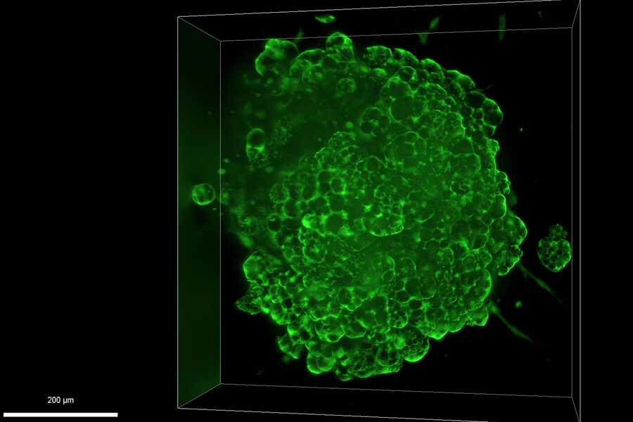
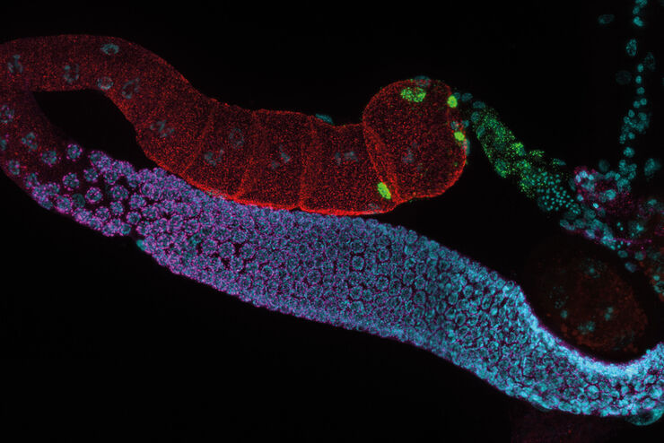
Murine esophageal organoids (DAPI, Integrin26-AF 488, SOX2-AF568) imaged with the THUNDER Imager 3D Cell Culture. Courtesy of Dr. F.T. Arroso Martins, Tamere University, Finland.

How to Get Deeper Insights into your Organoid and Spheroid Models

In this eBook, learn about key considerations for imaging 3D cultures, such as organoids and spheroids, and discover microscopy solutions to shed new insights into dynamic processes in 3D real-time
Brain organoid section (DAPI) acquired using THUNDER Imager Live Cell. Image courtesy of Janina Kaspar and Irene Santisteban, Schäfer Lab, TUM.

Imaging Organoid Models to Investigate Brain Health

Imaging human brain organoid models to study the phenotypes of specialized brain cells called microglia, and the potential applications of these organoid models in health and disease.
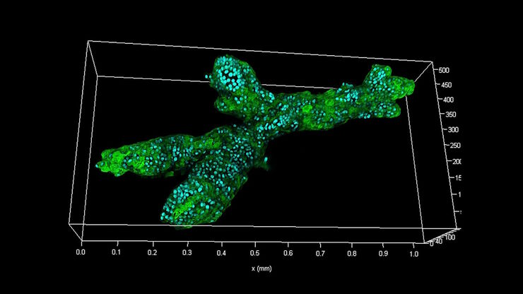
Branched organoid growing in collagen where the Nuclei are labeled blue. To detect the mechanosignaling process, the YAP1 is labeled green.

Examining Developmental Processes In Cancer Organoids

Interview: Prof. Bausch and Dr. Pastucha, Technical University of Munich, discuss using microscopy to study development of organoids, stem cells, and other relevant disease models for biomedical…
Krebszellen

Die Rolle des Eisenstoffwechsels bei der Krebsentwicklung

Der Eisenstoffwechsel spielt eine Rolle bei der Entstehung und dem Fortschreiten von Krebs und beeinflusst die Immunreaktion. Zu verstehen, wie Eisen Krebs und das Immunsystem beeinflusst, kann die…
Extended depth of field reconstruction image of a whole human pancreas islet showing fluorescence signals from insulin (green), glucagon (red), an IL17 cytokine (magenta), and nuclei (blue).

Understanding Better the Onset of Diabetes

This article shows how Interleukin-17 (IL-17) proinflammatory cytokine proteins in human pancreatic islets can be studied efficiently with a THUNDER Imager.

Harnessing Microfluidics to Maintain Cell Health During Live-Cell Imaging

VIDEO ON DEMAND - In this episode of MicaCam, we will use microfluidics to explore the effect of shear stress on cell morphology, examine the effect of nutrient replenishment on cellular growth during…

Effects of Clearing Media on Tissue Transparency and Shrinkage

This study comprehensively evaluates the effects of different clearing media on tissue transparency and shrinkage by comparing freshly dissected dipteran fly brains with their cleared equivalents.…

Fluorescence Lifetime-based Imaging Gallery

Confocal microscopy relies on the effective excitation of fluorescence probes and the efficient collection of photons emitted from the fluorescence process. One aspect of fluorescence is the emission…
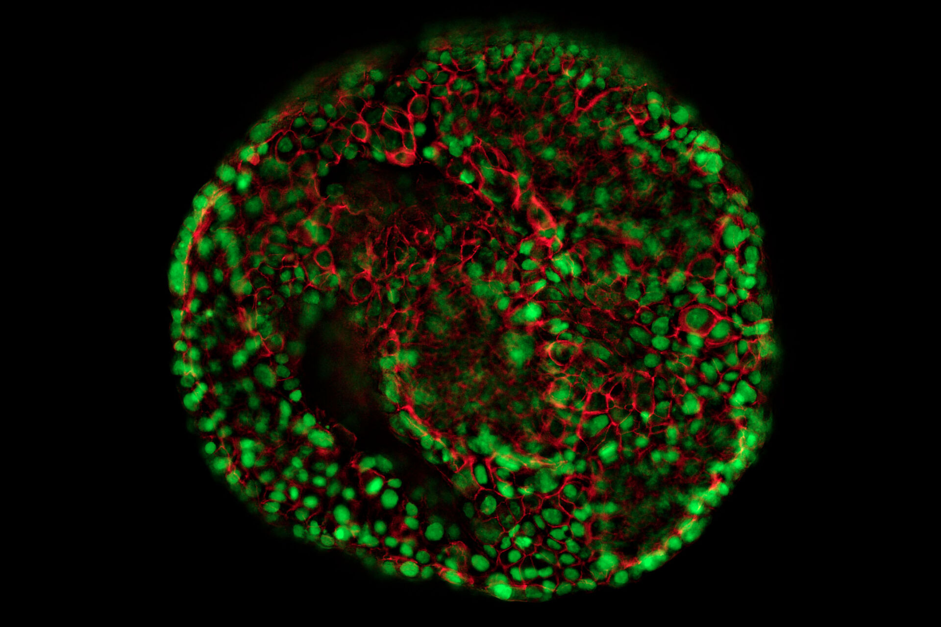
Images of a brain organoid derived from iPSCs acquired with a THUNDER Imager 3D Cell Culture. The cells were infected with the pAAV-hSyn-EGFP and pLX-hGFAP-mCherry virus. The image is the 36th plane cropped out of a 53 plane Z-stack volume. Shown are both the A) raw widefield image and B) the same image after Large Volume Computation Clearing (LVCC). Neurons are labeled in green and astrocytes in red.

“Brains-In-A-Dish” from Induced Pluripotent Stem Cells (iPSCs)

This article discusses the benefits of using the THUNDER technology for imaging inside 3D human cortical brain organoids. These organoids are derived from human induced pluripotent stem cells (iPSCs)…
Electroporated nerve cells (green), specific neuronal markers (magenta) and cell nuclei (white), computational cleared.

Into the Third Dimension with "Wow Effect"- Observe Cells in 3D and Real-Time

Life is fast, especially for a cell. As a rule, cells should be examined under physiological conditions which are as close as possible to their natural environment. New technologies offer tremendous…
Lung organoid taken at the "liquid-air interface" with a THUNDER Imager 3D Cell Culture. The cells originate from transgenic mice, so that the different fluorescence represents the degree of differentiation of the respective cell (superposition). The image acquisition was performed on day 21 after the start of the culture. Reference: P. Kanrai, MPI-HLR Bad Nauheim.

Observing 3D Cell Cultures During Development

3D cell cultures, such as organoids and spheroids, give insights into cells and their interactions with their microenvironment. These 3D cell cultures are playing an increasingly important role for…
Single-cell Huh-7D12 spheroid imaged using a Leica SP8 DLS microscope system. The spheroid was clarified using the Sucrose protocol.

A Quality Metric for the Systematic Evaluation of Clearing Protocols

3D multicellular spheroids are of interest for studying tumor behavior and evaluating the response of pharmacologically active agents, because they mimic the in vivo tumor environment better than…
Mouse lymphnode acquired with a THUNDER Imager 3D Cell Culture. Image courtesy of Dr. Selina Keppler, Munich, Germany.

Image Gallery: THUNDER Imager

To help you answer important scientific questions, THUNDER Imagers eliminate the out-of-focus blur that clouds the view of thick samples when using camera-based fluorescence microscopes. They achieve…
Elucidate cancer development on sub-cellular level by in-vivo like tumor spheroid models.

Improve 3D Cell Biology Workflow with Light Sheet Microscopy

Understanding the sub-cellular mechanisms in carcinogenesis is of crucial importance for cancer treatment. Popular cellular models comprise cancer cells grown as monolayers. But this approach…
Mammalian cell culure. Phase contrast and fluorescence image.

Introduction to Mammalian Cell Culture

Mammalian cell culture is one of the basic pillars of life sciences. Without the ability to grow cells in the lab, the fast progress in disciplines like cell biology, immunology, or cancer research…
Background image
Scroll to top