
About the interview
Key Interview Highlights
• How stem cells and organoids are contributing to disease research, such as mimicking cancerous processes.
• Why Mica is used to better understand spatiotemporal processes at the molecular or cellular scale.
• What the future holds for organoid research at TUM and its implications on biomedical research.
Interested to find out more about fast and stable live-cell imaging?
Transcript of the interview
Prof. Bausch
I am Andreas Bausch. I am the head of Cellular Biophysics at the Technical University of Munich, Director of the Center of Protein Assemblies, and founding Director of the Center for Organoid Systems.
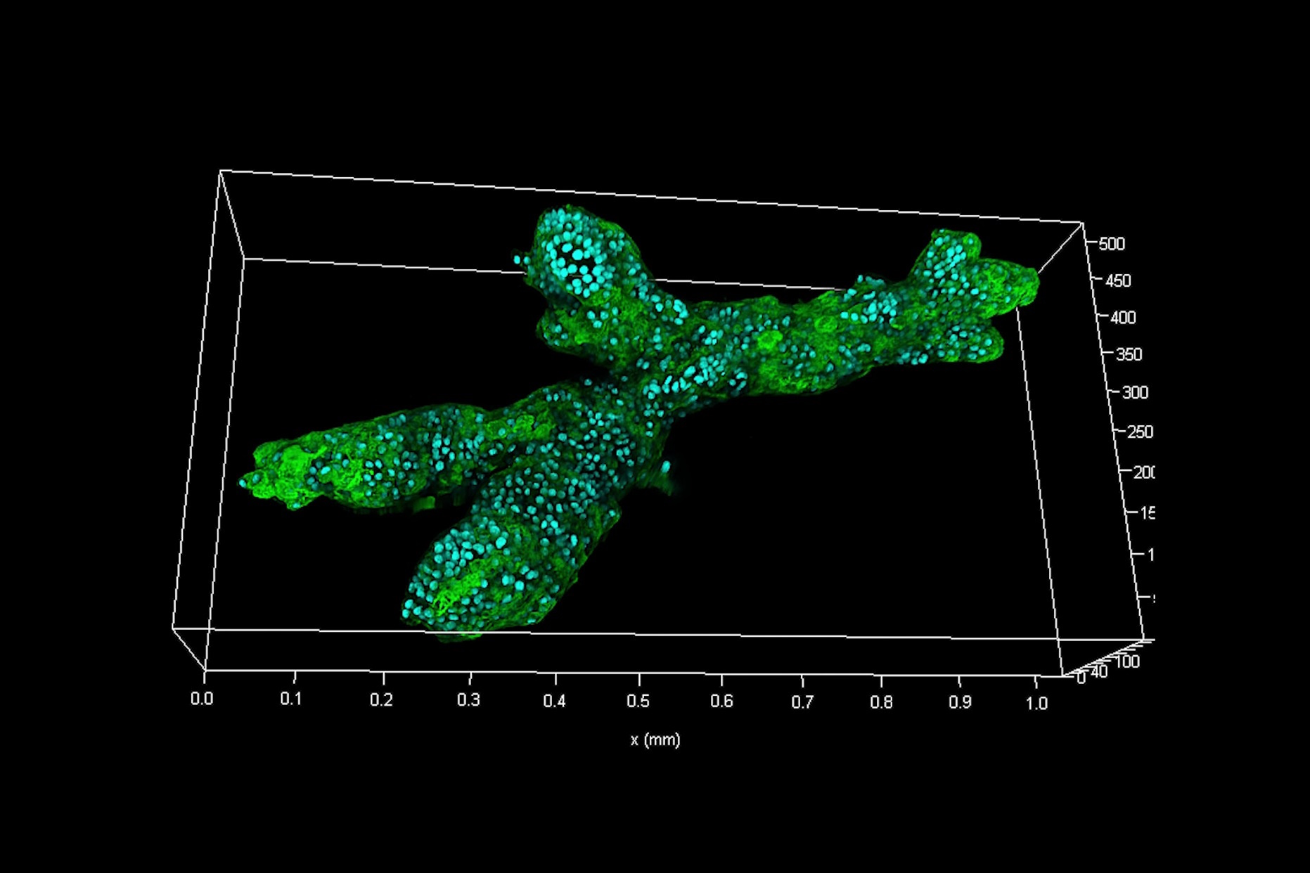
So, we looked recently at the development of mammary gland organoids. We have primary tissues of healthy human patients, and we use these cells to grow small mammary glands in this cell-culture tissue.
We are able to see all the development processes which lead then to structure formation in mammary glands. To unravel the physical basis of that, it is important to really observe the systems and really understand what kind of time-spatial processes are in them.
And for that microscopy is the natural choice of methods and light microscopy especially, because you have access to data of relevant length and time scales to understand how these processes happen at the molecular or cellular scale.
And then it becomes clear that's a kind of great tool to study health and disease and develop a nice disease model. Then in the cancer field you are able to mimic cancerous processes. These you must observe with light microscopy to understand what is going on.
Dr. Pastucha
My name is Anna Pastucha and I have worked in Professor Bausch’s Lab for a little more than one year.
One of the projects that I'm involved in is related to pancreatic cancer.
So, we work with pancreatic cancer organoids, which are very branched structures, and they grow in collagen. The collagen is a floating-in media so the whole system is very challenging for imaging. That's why we have the whole spectra of different types of microscopy systems. Here we have a standard confocal microscopy, FLIM, STELLARIS, and of course Mica, which combines standard epifluorescence microscopy and confocal microscopy.
So, whenever we find interesting structures and want to zoom in and have higher resolution, then, with basically one click, we can switch from the epifluorescence to confocal mode.
Additionally, we have one project which is related to stem-cell development and, of course, the stem cells require very stringent conditions.
That is why we use Mica for that, because it provides this very stable incubation chamber.
Also, what is pretty nice with Mica, and very important for us as well, is that it has a function which prevents photobleaching of samples and protects live cells from phototoxicity.
And for stem cells, it is very important that we keep the conditions very stable as well as that we don't kill them with too much light.
Prof. Bausch
I think it is pretty clear that these organoids have huge potential for biomedical research, from basic research up to applications like drug development, and the understanding and addressing of disease types.
And this is a huge field for which we have recently founded a Center for Organoid Systems, where we try to pull together biomedical engineers and basic scientists into one building, a new building where we try to really understand at all the different levels the applications for organoids. And that is something which has huge potential for future applications.
 and phalloidin (magenta), imaged using Viventis SCAPE; scale bar 50μm. Courtesy of Marina Cuenca and Heleen Jungen (Dayton lab), EMBL Barcelona.")


