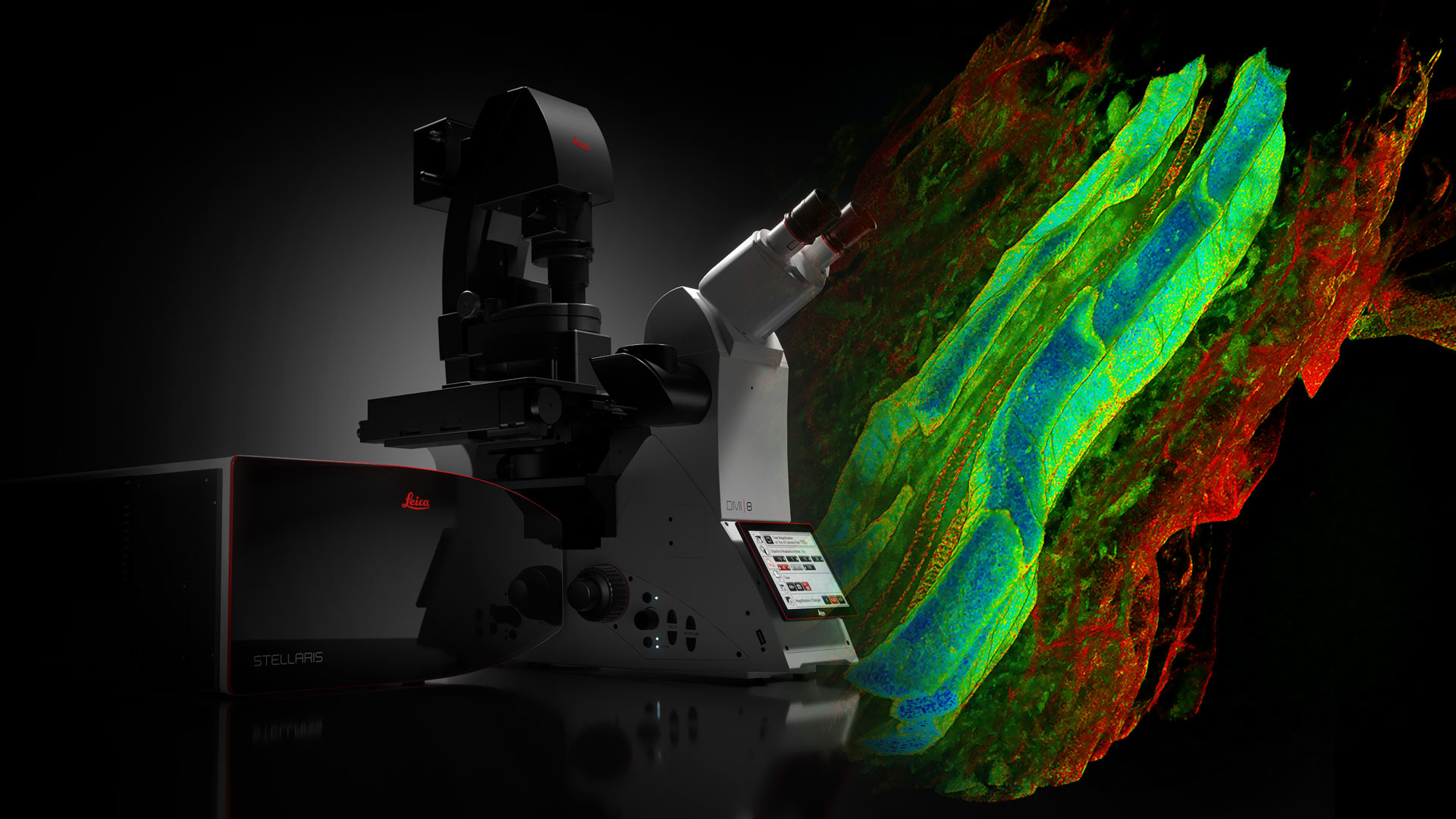
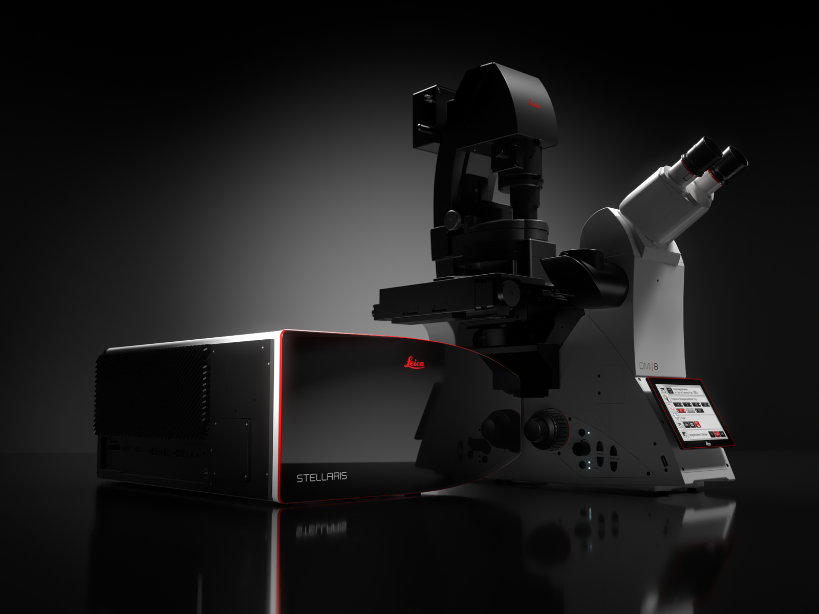
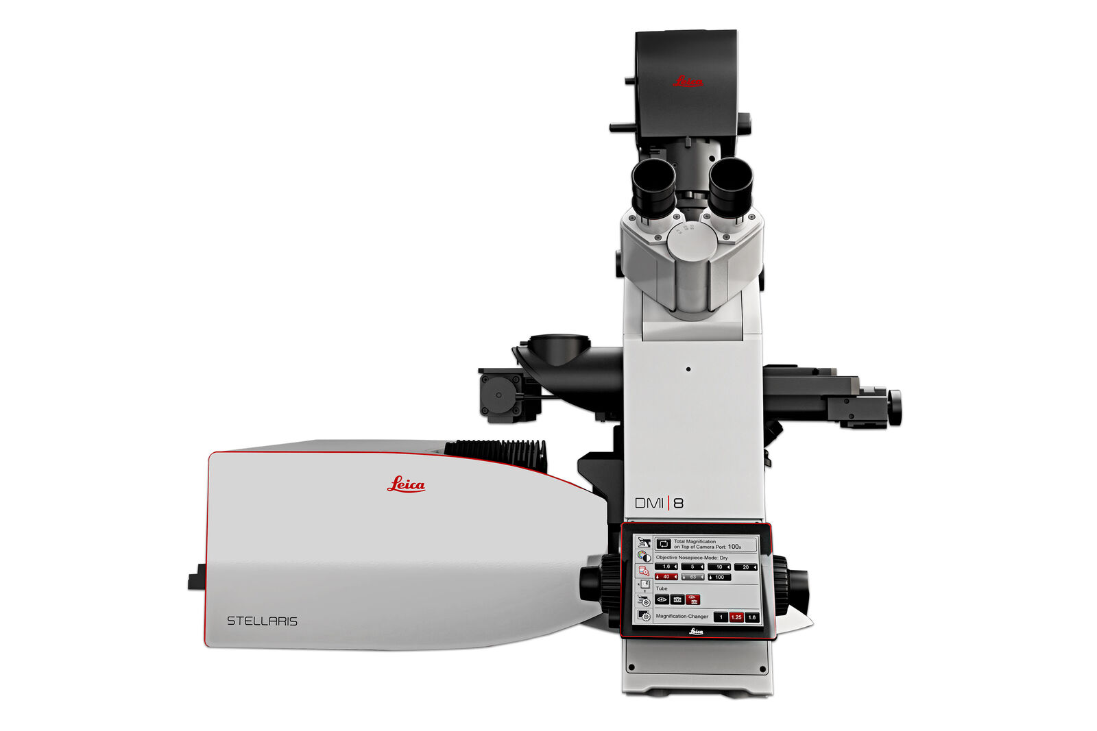
STELLARIS Plattform für konfokale Mikroskopie
STELLARIS der nächsten Generation ist eine Plattform für konfokale Mikroskopie, die auf Ihre Forschung zugeschnitten ist.
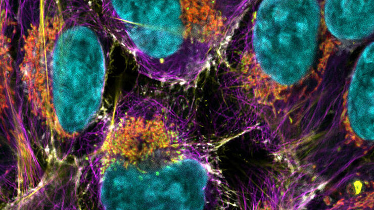
STELLARIS Konfokalmikroskope können mit allen Leica Modalitäten kombiniert werden, einschließlich FLIM, STED, MP, DLS und CRS. Die STELLARIS-Plattform für konfokale Mikroskopie der nächsten Generation ist Ihr schneller Weg zu ergebnisreicher Forschung. Power. Potential. Produktivität.
Das Potential, mehr zu entdecken
Die einzigartige TauSense-Technologie von STELLARIS ermöglicht Ihnen, aus jeder Probe zusätzliche Informationen zu gewinnen und dadurch den wissenschaftlichen Erkenntnisgewinn aus Ihrer Forschung zu steigern. TauSense besteht aus anwendungsorientierten Bildgebungstools auf der Grundlage der Fluoreszenzlebensdauer, mit denen Sie die Funktion von Molekülen im zellulären Kontext untersuchen können.
- Mit TauContrast haben Sie sofort Zugriff auf funktionelle Informationen wie Stoffwechselstatus, pH-Wert und Ionenkonzentration.
- TauGating verbessert die Qualität Ihrer Bilder, indem es unerwünschte Fluoreszenzbeiträge entfernt.
- TauSeparation hilft Ihnen, die Kombination von Fluoreszenzsignalen in Ihrem Experiment über die spektralen Optionen hinaus zu erweitern.
- TauInteraction ermöglicht die einfache Erkennung und Quantifizierung molekularer Wechselwirkungen (z. B. Protein-Protein-Wechselwirkung).
Zugriff auf relevante Daten
Die von Aivia unterstützte autonome Mikroskopie liefert Ihnen schneller hochwertige Ergebnisse.
- Der KI-basierte Workflow zur Erkennung seltener Ereignisse für die Biowissenschaften auf STELLARIS erkennt autonom bis zu 90 % der seltenen Ereignisse in biologischen Proben.
- Verkürzen Sie die Datenerfassungszeit drastisch um bis zu 70 %, da ausschließlich Objekte von Interesse identifiziert und erfasst werden. Speichern Sie nur das, was Sie brauchen, und sparen Sie wertvollen Speicherplatz auf Ihrer Festplatte.
- Weniger Zeit am Mikroskop verbringen: Der autonome Workflow zur Erkennung seltener Ereignisse benötigt nur einen Bruchteil der Zeit, die Sie normalerweise benötigen.
- Führen Sie mit der autonomen Mikroskopie von Aivia Experimente durch, die bisher aufgrund von Zeitbeschränkungen und Komplexität nicht möglich waren.
Die Produktivität, um mehr zu tun
Optimiertes Setup und Datenerfassung: ImageCompass, die intelligente Benutzeroberfläche von STELLARIS, bietet Benutzern eine einfache und intuitive Möglichkeit, auch besonders komplexe Experimente mit nur wenigen Klicks einzurichten.
- Einfach schnell: Hervorragende Bildqualität in Echtzeit und bei voller Geschwindigkeit mit der dynamischen Signalverstärkung (DSE) und Super-Auflösung LIGHTNING von Aivia.
- Intuitive Benutzeroberfläche: ImageCompass führt Sie vom Versuchsaufbau bis zur Aufnahme.
- Optimieren Sie Ihr Experiment: Tools wie der LAS X Navigator sind nahtlos integriert, um eine unkomplizierte Bildgebung zu ermöglichen.
STELLARIS mit WLL
STELLARIS ist das einzige Konfokalsystem mit integriertem Weißlichtlaser (WLL), kombiniert mit unserem proprietären akustooptischen Strahlteiler (AOBS) und den neuen Power HyD-Detektoren. Zusammen mit der einzigartigen TauSense-Technologie setzt STELLARIS einen neuen Standard hinsichtlich der Bildqualität und der Menge an gewonnenen Informationen.
STELLARIS mit Weißlichtlaser kann mit FAst Lifetime CONtrast (FALCON), STED, Deep In Vivo Explorer (DIVE), Digital Light Sheet (DLS), Cryo und CARS kombiniert werden. STELLARIS maximiert das Leistungsvermögen dieser Modalitäten und gibt Ihnen die Power und das Potential, neue Standards in der Forschung zu setzen.
STELLARIS
STELLARIS bietet Spektralerkennung, Photon-Counting und konfokale Superauflösung.
Die konfokale Mehrkanal-Bildgebung ist dank der intelligenten Benutzeroberfläche ImageCompass einfach durchführbar, da Sie intuitiv durch die Einrichtung und Erfassung Ihres Experiments geführt werden.
Die Power, mehr zu sehen
Die Detektoren der Power HyD-Familie bieten eine höhere Photonenerkennungseffizienz (PDE)*, extrem geringes Dunkelrauschen und eine empfindliche Spektralerkennung von 410 bis 850 nm.
- Bessere Bildqualität: Perfekte Kombination aus Helligkeit, Auflösung und Kontrast.
- Vollständige spektrale Freiheit: Mit unseren Weißlichtlasern der nächsten Generation können Sie bis zu 8 einzelne Anregungslinien aus dem gesamten Spektrum gleichzeitig nutzen. Bilden Sie mehr Fluorophorkombinationen ab als mit jeder anderen konfokalen Plattform. Verwenden Sie mehr Labels geichzeitig und erweitern Sie Ihre Optionen in den NIR-Bereich.
- Schonende Bildgebung lebender Zellen: Bewahren Sie die Unversehrtheit Ihrer Probe und bilden Sie sie über einen längeren Zeitraum ab dank der effizienten Signalerfassung bei niedrigsten erforderlichen Beleuchtungsstärken.
* Zweifach höhere Photonendetektionseffizienz (PDE) im Vergleich zu herkömmlichen Multi-Alkali-Photomultiplier-Röhren (PMT) und dreifach höhere PDE im erweiterten Rotbereich.