is mobile? false
Produkte
Produkte
Unsere High-Tech-Präzisionssysteme für die Analyse von Mikrostrukturen entwickeln wir mit dem Anwender für den Anwender. In unserem Produktportfolio finden Sie Systemlösungen im Bereich Life Science einschließlich Biotechnologie und Medizin sowie Werkstoffwissenschaft und industrielle Qualitätskontrolle.
Produkt Kategorien Show subnavigation
Produkte
Präzisionsmikroskopieprodukte für die Analyse von Mikrostrukturen. Mikroskoplösungen für Life Science, Medizin, Forschung und Entwicklung sowie industrielle Qualitätssicherung.
Veranstaltungen Show subnavigation
Werfen Sie einen Blick auf alle unsere anstehenden Konferenzen, Kongresse, Messen, Webinare und Workshops und besuchen Sie uns bei einer unserer nächsten Veranstaltungen!
Anwendungen Show subnavigation
Leica Science Lab Articles Show subnavigation
Lesen Sie unsere neuesten Artikel
Das Wissensportal von Leica Microsystems bietet Ihnen Wissens- und Lehrmaterial zu den Themen der Mikroskopie. Die Inhalte sind so konzipiert, dass sie Einsteiger, erfahrene Praktiker und Wissenschaftler gleichermaßen bei ihrem alltäglichen Vorgehen und Experimenten unterstützen.
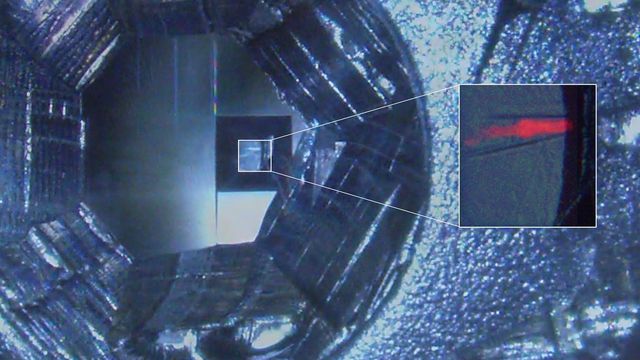
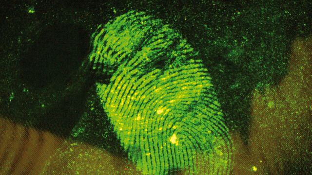
How Fluorescence Guides Sectioning of Resin-embedded EM Samples
Electron microscopes, including transmission electron microscopes (TEM) and scanning electron microscopes (SEM), are widely utilized to gain detailed structural information about biological samples or non-living materials. Ultramicrotomy is the preferred technique for producing ultrathin sections, less than 100 nm thick for TEM/SEM analysis. During sample preparation small sample pieces are embedded in epoxy or acrylic resin, excess resin is trimmed away, and the specimen is sliced into ultrathin sections (50 nm - 100 nm) using a glass or diamond knife.
Coherent Raman Scattering Microscopy Publication List
CRS (Coherent Raman Scattering) microscopy is an umbrella term for label-free methods that image biological structures by exploiting the characteristic, intrinsic vibrational contrast of their molecules. The two most important CRS techniques are Coherent Anti-Stokes Raman Scattering (CARS) and Stimulated Raman Scattering (SRS). The biochemical image contrast of CRS is in many ways complementary to the molecular contrast obtained in fluorescence microscopy. A second crucial advantage of these methods is that they preserve the specimen/sample in a near pristine state. This reference list presents current and basic papers on CRS microscopy.
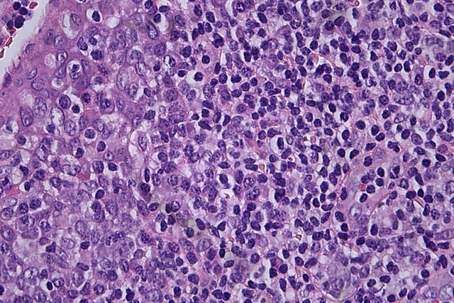
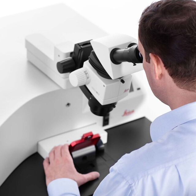
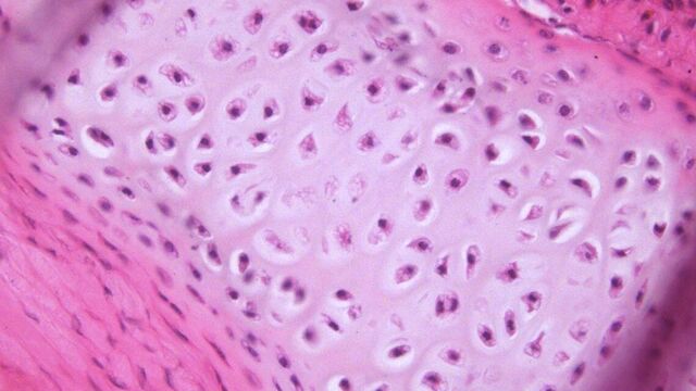
Clinical Microscopy: Considerations on Camera Selection
The need for images in pathology laboratories has significantly increased over the past few years, be it in histopathology, cytology, hematology, clinical microbiology, or other applications. They serve many purposes on top of the documentation of the diagnosis. Yet, the view through the eyepieces and the image are different in nature, the one is an optical image, the other a digital image. Looking at a few aspects of this process that are related cameras will help you make sure you can obtain the images with all the detail and color fidelity you need.
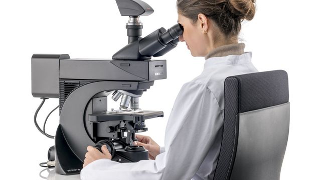
Factors to Consider when Selecting Clinical Microscopes
What matters if you would like to purchase a clinical microscope? Learn how to arrive at the best buying decision from our Science Lab Article.
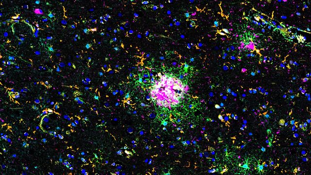
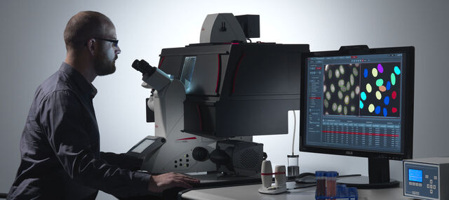
Explore Alzheimer's Spatial Proteome with Big Data
Alzheimer's disease, a genetic and sporadic neurodegenerative condition, leads to cognitive decline in mid to late life, marked by β-amyloid plaques and tau tangles. With limited treatment options, new investigative strategies are crucial. The Cell DIVE multiplexed imaging solution allows examination of Alzheimer's brain tissue, potentially uncovering new research avenues. Here, we showcase the Cell DIVE image viewer, enabling users to access the full Alzheimer's multiplexed dataset directly in their browser.
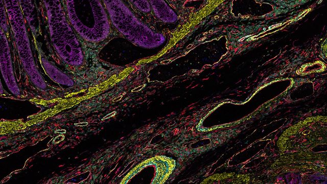
Uncover the Hidden Complexity of Colon Cancer with Big Data
Colorectal cancer poses a significant health burden. While surgery is effective initially, some patients develop recurrent secondary disease with poor prognosis, necessitating advanced therapies like immunotherapies. Spatial biology approaches, such as multiplexed imaging with Cell DIVE, can provide crucial insights for developing novel treatments. Access the full Cell DIVE dataset in your browser to explore these findings further through the Minerva image viewer.
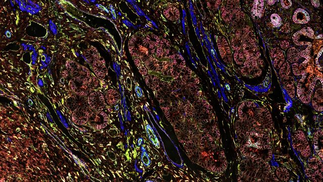
Dive into Pancreatic Cancer Research with Big Data
Pancreatic cancer, with a mortality rate near 40%, is challenging to treat due to its proximity to major organs. This story explores the complex biology of pancreatic ductal adenocarcinoma (PDAC), examining molecular and spatial determinants of tumor aggression in metabolism, apoptosis, and immunity. Access the full Cell DIVE dataset in your browser to delve deeper into these findings.
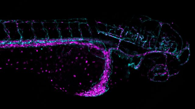
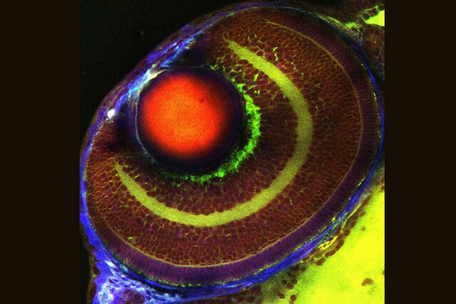
Overcoming Challenges with Microscopy when Imaging Moving Zebrafish Larvae
Zebrafish is a valuable model organism with many beneficial traits. However, imaging a full organism poses challenges as it is not stationary. Here, this case study shows how zebrafish larvae can be imaged during stationary periods and easily relocated after movement. The seamlessly integrated widefield and confocal capability of Mica is leveraged to capture fast events, like the heartbeat, with virtually no out-of-focus background noise which is inherent to standard widefield systems.
