Software per imaging al microscopio

Software per imaging al microscopio

Software per imaging al microscopio

Il software per imaging al microscopio di Leica Microsystems, combina un microscopio ottico, una fotocamera digitale e tutti gli accessori in un'unica soluzione completamente integrata.

Leggi i nostri articoli più recenti relativi al software per imaging al microscopio

Il portale informativo di Leica Microsystems offre materiale didattico e di ricerca scientifica su vari temi della microscopia. Il contenuto è stato progettato per aiutare i principianti, i professionisti e gli scienziati nel lavoro quotidiano e nella ricerca avanzata.

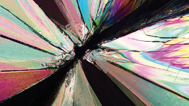
The Polarization Microscopy Principle

Polarization microscopy is routinely used in the material and earth sciences to identify materials and minerals on the basis of their characteristic refractive properties and colors. In biology, polarization microscopes are commonly used for identification of birefringent structures, like crystals, or for imaging of cellulose in the walls of plant cells and starch grains. This article gives an overview of the basic principles of polarization microscopy.

Super-Resolution Microscopy Image Gallery

Due to the diffraction limit of light, traditional confocal microscopy cannot resolve structures below ~240 nm. Super-resolution microscopy techniques, such as STED, PALM or STORM or some deconvolution processing methods, are used when enhanced resolution is needed to study structures and molecular events below the diffraction limit scale.

Extended Live-cell Imaging at Nanoscale Resolution

Extended live-cell imaging with TauSTED Xtend. Combined spatial and lifetime information allow super-resolution microscopy at extremely low light dose.
Mar 05, 2024
Webinar
STED

Studying Virus Replication with Fluorescence Microscopy

The results from research on SARS-CoV-2 virus replication kinetics, adaption capabilities, and cytopathology in Vero E6 cells, done with the help of fluorescence microscopy, are described in this article.

Epi-Illumination Fluorescence and Reflection-Contrast Microscopy

This article discusses the development of epi-illumination and reflection contrast for fluorescence microscopy concerning life-science applications. Much was done by the Ploem research group collaborating with the company Leitz.

Going Beyond Deconvolution

Widefield fluorescence microscopy is often used to visualize structures in life science specimens and obtain useful information. With the use of fluorescent proteins or dyes, discrete specimen components are marked in a highly specific manner. To fully understand a structure, visualizing it in 3 dimensions can be necessary, but certain challenges are faced when doing so with microscopy.

AI Microscopy Image Analysis – An Introduction

Artificial intelligence-guided microscopy image analysis and visualization is a powerful tool for data-driven scientific discovery. AI can help researchers tackle challenging imaging applications, allowing them to extract more information from their images.

The Potential of Coherent Raman Scattering Microscopy at a Glance

Coherent Raman scattering microscopy (CRS) is a powerful approach for label-free, chemically specific imaging. It is based on the characteristic intrinsic vibrational contrast of molecules in the sample. CRS provides high-resolution (sub-cellular level) and dynamic (up to video rate) information on the biochemical composition and metabolic processes in cells, tissues, and intact model organisms. It also enables imaging of small molecules without perturbing their function. This information is highly synergistic with the molecular contrast provided by fluorescence microscopy. Unsurprisingly, CRS is finding a growing number of applications in fields like neurodegenerative disease, cancer, 3D biology, stem cell and developmental biology, and pharmacology.

Simplifying Complex Fluorescence Multiwell Plate Assays

Apoptosis, or programmed cell death, occurs during organism embryo development to eliminate unwanted cells and during healing in adults to rid the body of damaged cells and help prevent cancer. Caspase assay experiments with multiwell plates allow researchers to study the early phases of apoptosis. In this article, we show how Mica can be used in this key application with fluorescence multiwell plate assays to provide 100% spatiotemporal correlation of data and minimize crosstalk .

Efficient Long-term Time-lapse Microscopy

When doing time-lapse microscopy experiments with spheroids, there are certain challenges which can arise. As the experiments can last for several days, prolonged sample survival must be achieved which requires that near physiological conditions are ensured. The long-term time-lapse study described in this article involves the use of Mica - the world’s first Microhub, to investigate the formation of spheroids from U343 and MDCK cells. Growing spheroids requires optimal conditions to ensure undisturbed cell cycles and proliferation.

Multicolor 4D Super Resolution Light Sheet Microscopy

The AI Microscopy Symposium offers a unique forum for discussing the latest AI-based technologies and tools in the field of microscopy and biomedical imaging. In this scientific presentation, Yuxuan Zhao demonstrates how 3D imaging of organelles within live cells can be improved using a progressive deep learning strategy combined with a dual-ring-modulated SPIM design.

Hyperplex Cancer Tissue Analysis at Single Cell Level with Cell DIVE

The ability to study how lymphoma cell heterogeneity is influenced by the cells’ response to their microenvironment, especially at the mutational, transcriptomic, and protein levels. Protein expression studies offer the most relevant information about the nature of cellular interactions and protein expression levels. A hyperplexed workflow can be applied for studying multiple proteins from the same cancer tissue.

How to Prepare your Specimen for Immunofluorescence Microscopy

Immunofluorescence (IF) is a powerful method for visualizing intracellular processes, conditions and structures. IF preparations can be analyzed by various microscopy techniques (e.g. CLSM, Epifluorescence, TIRF, GSDIM), depending on the application or the researcher’s interest. Meanwhile, IF has become indispensable for a large number of research groups which have at least access to a simple fluorescence microscope.

Live-Cell Imaging Techniques

The understanding of complex and/or fast cellular dynamics is an important step for exploring biological processes. Therefore, today’s life science research is increasingly focused on dynamic processes like cell migration, morphological changes of cells, organs, or whole animals, and real-time physiological (e.g., changes of intracellular ion composition) events in living specimens. One approach to address these challenging demands is to employ certain optical methods that are collectively called live-cell imaging.

Fluorescent Dyes

A basic principle in fluorescence microscopy is the highly specific visualization of cellular components with the help of a fluorescent agent. This can be a fluorescent protein – for example GFP – genetically linked to the protein of interest. If cloning is impossible – for instance in histologic samples – techniques such as immunofluorescence staining are used to visualize the protein of interest.

The AI-Powered Pixel Classifier

Achieving reproducible results manually requires expertise and is tedious work. But now there is a way to overcome these challenges by speeding up this analysis to extract the real value of the image and gain insights. The artificial-intelligence-powered pixel classifier provides reproducible segmentation results fast and overcomes human action. It delivers more robust results compared to classical function-based automation.

Tutorial sul software LAS X

Vuoi saperne di più sul software LAS X o hai bisogno di consigli su come utilizzarlo?

Visita il nostro canale YouTube!

Sono presenti numerosi video con utili tutorial e suggerimenti su come utilizzare LAS X nei microscopi biologici, industriali e confocali:

Leica Microsystems Tutorials

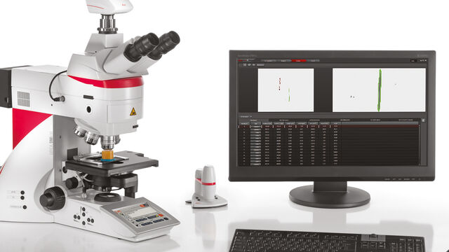
Una soluzione completa

Grazie all'interfaccia utente intuitiva e alla navigazione semplificata, il nostro software per imaging al microscopio guida l'utente attraverso un flusso di lavoro personalizzato, sia per l'acquisizione rapida che per l'analisi professionale delle immagini. La gamma di moduli specifici consente di configurare il microscopio come strumento ad alte prestazioni dedicato a tutte le applicazioni.

La più recente piattaforma software, LAS X, include tutte le soluzioni per microscopia sia per applicazioni nei settori industriali che nelle scienze biologiche, garantendo la massima flessibilità. La precedente versione, Leica Application Suite, continua ad essere supportata.

Caratteristiche principali

  • Configurazione e controllo dei microscopi e delle fotocamere digitali in modo completamente integrato.
  • Gli strumenti di annotazione di base consentono di aggiungere immagini e valori della calibrazione alle immagini.
  • Le regolazioni manuale e automatica dell’esposizione consentono di ottimizzare le condizioni di imaging.
  • Una galleria di miniature delle immagini acquisite, permette di riesaminarle in modo rapido e semplice.
  • Immagini calibrate automaticamente utilizzando i dati provenienti dai microscopi e fdalle fotocamere Leica; una scala di calibrazione indica le dimensioni dell'immagine.
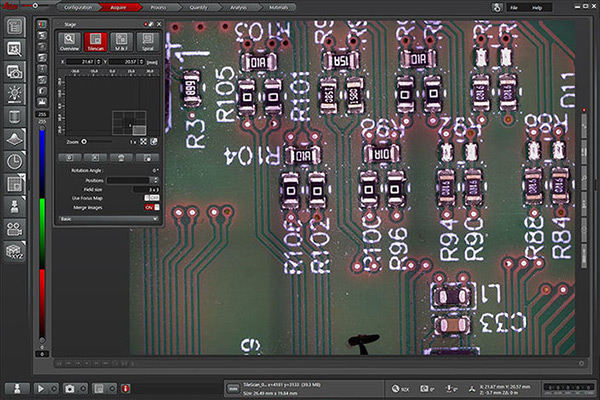
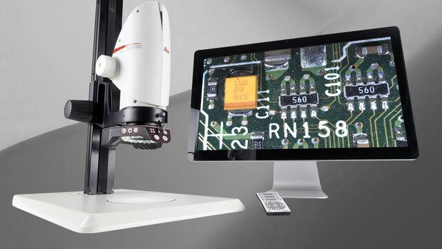
     

LAS X Industry

  • Osserva il campione a schermo intero
  • Esegui le misurazioni in modo semplice e flessibile
  • Migliora l'imaging con la composizione sugli assi X, Y e Z 
  • Acquisisci e richiama i risultati in modo affidabile e riproducibile
  • Personalizza la stampa dei report in base alle tue esigenze
     

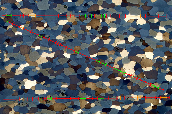
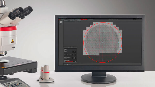
Moduli LAS X Materials Science

  • Consente agli utenti di eseguire rapidamente l'analisi strutturale dell'acciaio e di altri materiali.
  • Consente l'analisi professionale di fasi multiple e di componenti di microstrutture.
  • Sviluppato appositamente per combinare misurazioni accurate di oggetti e aree con un'elevata flessibilità per personalizzare le analisi automatizzate in base alle esigenze specifiche dell'utente.
  • Offre un miglioramento significativo dell'ergonomia e aiuta gli utenti a mantenere una precisione nelle misurazioni costante nel tempo.
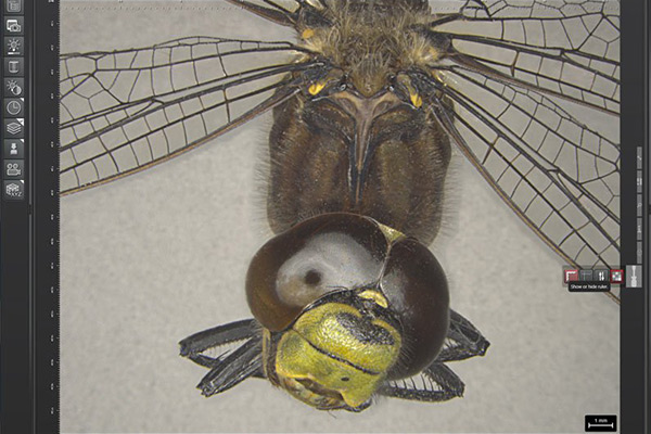
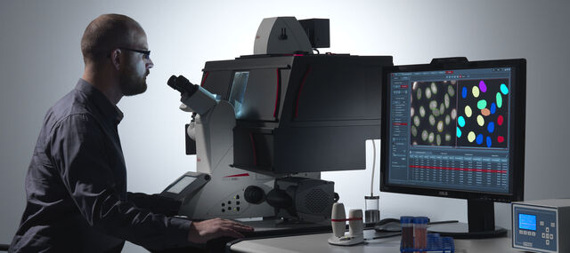
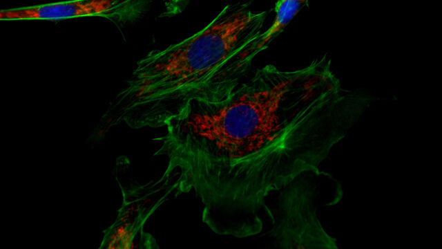
     

LAS X Life Science

  • Integra gli strumenti per la microscopia confocale, ottici, stereo, a super risoluzione e light-sheet di Leica Microsystems.
  • Crea panoramiche rapide e identifica immediatamente i dettagli principali con LAS X Navigator, il GPS delle cellule
  • Le attività di imaging diventano intuitive e consentono di concentrarsi sulla ricerca
     
Scroll to top