THUNDER Imager Cell Spinning Disk
倒立顕微鏡
光学顕微鏡
製品紹介
Home
Leica Microsystems
THUNDER Imager Cell Spinning Disk
Clarity Through Synergy
最新の記事を読む
, Tropomyosin (cardiomyocytes, red) and GFP (primordial cardiac layer, green).")
A Guide to Fluorescence Microscopy
Fluorescence microscopy uses the ability of fluorophores, dyes, or fluorescent proteins to emit light of a specific wavelength after being excited with light of a shorter wavelength. Biomolecules can…
 and astrocytes (green) in a cortical spheroid derived from human induced pluripotent stem cells.")
Guide to Live-Cell Imaging
For a wide range of applications in various research fields of life science, live-cell imaging is an indispensable tool for visualizing cells in a state as close to in vivo, i.e. living and active, as…

Factors to Consider When Selecting a Research Microscope
An optical microscope is often one of the central devices in a life-science research lab. It can be used for various applications which shed light on many scientific questions. Thereby the…
 and tubulin (magenta), acquired using Viventis Deep. Courtesy of Akanksha Jain, Treutlein Lab ETH-DBSSE Basel (Switzerland).")
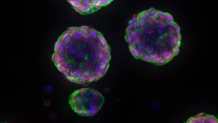
Faster & Deeper Insights into Organoid and Spheroid Models
Gain deeper, more translatable, insights into organoid and spheroid models for drug discovery and disease research by overcoming key imaging challenges. In this eBook, explore advanced microscopy…

A Guide to C. elegans Research – Working with Nematodes
Efficient microscopy techniques for C. elegans research are outlined in this guide. As a widely used model organism with about 70% gene homology to humans, the nematode Caenorhabditis elegans (also…

Boston and San Francisco Innovation Hubs
Boston and San Francisco Innovation Hubs are here to help you advance scientific discovery. We provide researchers access to state-of-the-art microscope technology and expert guidance. Located in the…

Unlocking the Secrets of Organoid Models in Biomedical Research
Get ready to delve deeper into the world of organoids and 3D models, which are essential tools for advancing our understanding of human health. Navigating these complex structures and obtaining clear…

Total Internal Reflection Fluorescence (TIRF) Microscopy
Total internal reflection fluorescence (TIRF) is a special technique in fluorescence microscopy developed by Daniel Axelrod at the University of Michigan, Ann Arbor in the early 1980s. TIRF microscopy…

Applications of TIRF Microscopy in Life Science Research
The special feature of TIRF microscopy is the employment of an evanescent field for fluorophore excitation. Unlike standard widefield fluorescence illumination procedures with arc lamps, LEDs or…
バイオファーマ
ライカのソリューションは、バイオ医薬品業界において、創薬の促進、細胞解析の強化、規制を満たすデータの完全性をサポートします。

Advanced Tissue Imaging & Analysis
Gain insights into tissue structure and function to improve your understanding of spatial biology and disease mechanisms with advanced imaging solutions from Leica Microsystems.
Fields of Application
オルガノイドと3D細胞培養
ライフサイエンス研究で近年最も目覚ましい発展の一つは、オルガノイド、スフェロイド、生体機能チップモデルなどの3D細胞培養システムの開発です。 3D細胞培養は、細胞が成長し、全3次元で周囲と相互作用できる人工環境です。 これらの条件は、生体内条件に似ています。
バイオファーマ
ライカのソリューションは、バイオ医薬品業界において、創薬の促進、細胞解析の強化、規制を満たすデータの完全性をサポートします。
