SR GSD 3D 3D-Lokalisationsmikroskop
The Evolution of Resolution
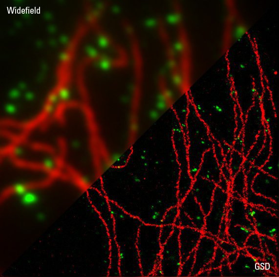
PtK2 cells with Alexa647 stained microtubules in red and Alexa488 stained clathrin in green
Courtesy: Prof. Dr. Stefan Hell, Max-Planck-Institute for Biophysical Chemistry, Göttingen, Germany
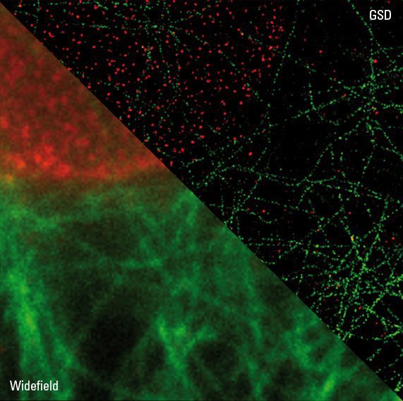
Ptk2-cells. NPC-staining: anti-NUP153/Alexa FLUOR 532 Microtubule-staining: anti-β-tubulin/Alexa FLUOR 488
Courtesy: Wernher Fouquet, Leica Microsystems in collaboration with Anna Szymborsak and Jan Ellenberg, EMBL, Heidelberg, Germany
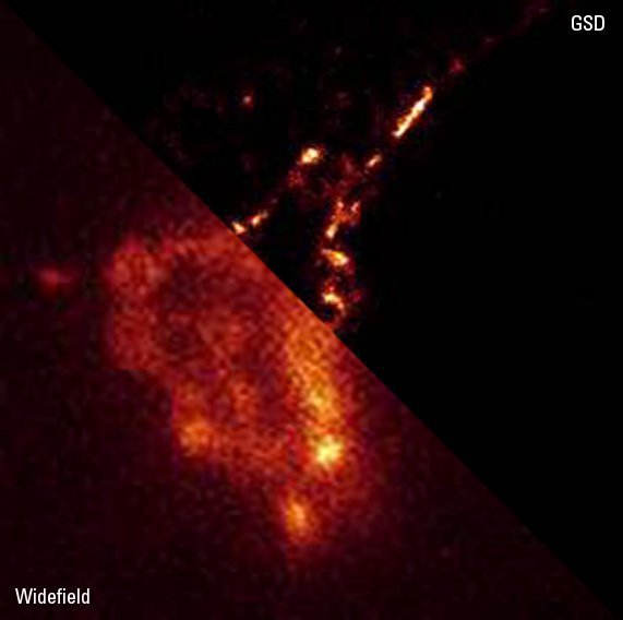
Golgi body, B16 (Mouse melanoma cell line), Golgi targeting signal of β-1,4-galactosyltransferase, fused to EYFP
Courtesy: Dr. Yasushi Okada, Department of Cell Biology and Anatomy, Graduate School of Medicine, University of Tokyo, Japan
Vimentin Filaments
3D reconstruction of a corner of a human endothelial cell (Huvec), stained for Vimentin with Alexa 647 Ab. Medium: gloxy-like buffer. Courtesy of K. Jalink and L. Nahidi Azar, Amsterdam, The Netherlands.
Neuron synapse
3D reconstruction of a synapse. Double staining of post- and presynaptic proteins Homer /Alexa647 (red) and Bassoon/ Alexa532 (green). Embedding media: MEA. Courtesy of Dr. W. Zuschratter, Dr. O. Kobler, Leipniz Institute for Neurobiology Magdeburg, Germany
Mitochondrial ATP-Synthase
Comparison of a 3D reconstruction of GSD and wide-field images. Mitochondrial ATP-Synthase in MDCK cells was stained with anti-ATP-2B/Alexa647 and four image stacks each 800 nm in thickness were merged. The color code indicates the total height of the image. Courtesy of R. Jacob, Marburg, Germany…
Vesicle Protein
MDCK cells stably transfected with a vesicle membrane marker protein fused to YFP. Recombinant Protein of interest coupled with Alexa 647 was endocytosed from the apical membrane of the cell. After internalization the cells were fixed and stained with an anti-EGFP-antibody/Atto 532 to increase the…
3D reconstruction of neurons
3D reconstruction of neurons, double stained for postsynaptic protein Homer with Alexa647® (red) and presynaptic protein Bassoon with Alexa532® (green). Embedding medium: MEA. Courtesy of Dr. W. Zuschratter, Dr. O. Kobler, Leipniz Institute for Neurobiology Magdeburg, Germany.
3D reconstruction of the synaptonemal complex protein 3 (SYCP3)
3D reconstruction of the synaptonemal complex protein 3 (SYCP3) stained with Alexa 647. Medium: 50 mM MEA in PBS. Five image stacks each 800 nm in thickness were merged. Courtesy of M. Lessard, Maine, USA (sample preparation).


