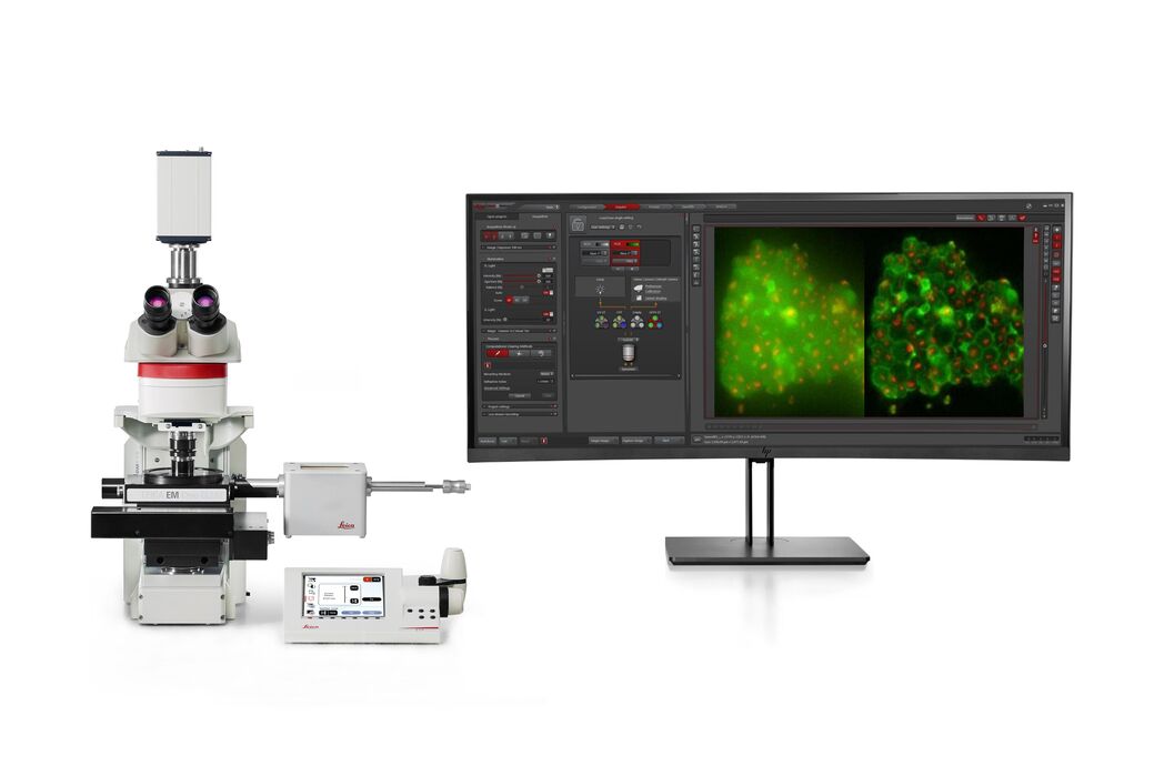
THUNDER Imager EM Cryo CLEM Kryo-Lichtmikroskop

Tiefgehendes Verständnis der zellulären Strukturbiologie

Lesen Sie unsere neuesten Artikel

How to Successfully Perform Live-cell CLEM

The Leica Nano workflow provides a streamlined live-cell CLEM solution for getting insight bout structural changes of cellular components over time. Besides the technical handling described in the…

How to Successfully Implement Coral Life

The live-cell  CLEM workflow allows you to capture dynamic information related to a relevant biological process as it happens and put these observations into their ultrastructural context. The Leica…

Advancing Cellular Ultrastructure Research

Freeze-fracture and freeze-etching are useful tools for studying flexible membrane-associated structures such as tight junctions or the enteric glycocalyx. Freeze-fracture and etching are two…

The Cryo-CLEM Journey

This article describes the Cryo-CLEM technology and the benefits it can provide for scientists. Additionally, some scientific publications are highlighted. Recent developments in cryo electron…
3D reconstruction of an intercellular bridge in a C. elegans embryo

Download EM Workflow Solutions Booklet

This publication is a compilation of appropriate workflows for the most frequently used sample preparation methods, like Correlative Methodologies, Optogenetics & Electro-Physiology, Surface Analysis,…

Exploring the Structure and Life Cycle of Viruses

The SARS-CoV-2 outbreak started in late December 2019 and has since reached a global pandemic, leading to a worldwide battle against COVID-19. The ever-evolving electron microscopy methods offer a…
Cryo FIB lamella - Overlay of SEM and confocal fluorescence image. Target structure in yeast cells (nuclear pore proteine Nup159-Atg8-split Venus, red) marked by an arrow. Scale bar: 5 µm. Alegretti et al.,  Nature 586, 796-800 (2020).

Targeting Active Recycling Nuclear Pore Complexes using Cryo Confocal Microscopy

In this article, how cryo light microscopy and, in particular cryo confocal microscopy, is used to improve the reliability of cryo EM workflows is described. The quality of the EM grids and samples is…

Advancing Cell Biology with Cryo-Correlative Microscopy

Correlative light and electron microscopy (CLEM) advances biological discoveries by merging different microscopes and imaging modalities to study systems in 4D. Combining fluorescence microscopy with…

Workflows and Instrumentation for Cryo-electron Microscopy

Cryo-electron microscopy is an increasingly popular modality to study the structures of macromolecular complexes and has enabled numerous new insights in cell biology. In recent years, cryo-electron…
Mouse retina was fixed and stained by following reagents: anti-CD31 antibody (green): Endothelia cells, IsoB4 (red): Blood vessels, and microglia anti-GFAP antibody (blue): Astrocytes Sample courtesy by Jeremy Burton, PhD and Jiyeon Lee, PhD, Genentech Inc., South San Francisco, USA. Imaged by Olga Davydenko, PhD (Leica). Imaged with a THUNDER Imager 3D Cell Culture.

An Introduction to Computational Clearing

Many software packages include background subtraction algorithms to enhance the contrast of features in the image by reducing background noise. The most common methods used to remove background noise…

Crystal Clear Cryo Light-microscopy Images

This article describes how computational clearing of cryo light microscopy images improves the identification of cellular targets for cryo electron-microscopy.

Real Time Images of 3D Specimens with Sharp Contrast Free of Haze

THUNDER Imagers deliver in real time images of 3D specimens with sharp contrast, free of the haze or out-of-focus blur typical of widefield systems. They can even image clearly places deep inside a…

Anwendungsbereiche

Fluoreszenz

Die Fluoreszenz ist eines der am häufigsten verwendeten physikalischen Phänomene in der biologischen und analytischen Mikroskopie, vor allem wegen ihrer hohen Empfindlichkeit und Spezifität. Erfahren…

Krebsforschung

Krebs ist eine komplexe und heterogene Krankheit, die durch Zellen verursacht wird, denen die Wachstumsregulation fehlt. Genetische und epigenetische Veränderungen in einer Zelle oder einer Gruppe von…

Zellbiologie

Erfahren Sie, wie Sie mit Bildgebungslösungen von Leica Microsystems das Beste aus Ihrer zellbiologischen Forschung herausholen und Ihre Entdeckungen erweitern können.

Virologie

Liegt Ihr Forschungsschwerpunkt auf Virusinfektionen und -krankheiten? Erfahren Sie, wie Sie mit Lösungen für Bildgebung und Probenvorbereitung von Leica Microsystems mehr Erkenntnisse in der…

Modellorganismen in der Forschung

Modellorganismen sind Spezies, mit denen Forscher bestimmte biologische Vorgänge untersuchen. Sie haben genetische Ähnlichkeiten mit Menschen und werden häufig in Forschungsbereichen wie Genetik,…

Kryo-Elektronen-Tomographie

Cryo-electron tomography (CryoET) is used to resolve biomolecules within their cellular environment down to an unprecedented resolution below one nanometer.

Fortgeschrittene Mikroskopietechniken

Moderne Mikroskopietechniken umfassen sowohl hochauflösende als auch superauflösende Bildgebungsverfahren. Diese Techniken werden in erster Linie eingesetzt, um biologische Vorgänge mit extrem hoher…

Korrelative Licht- und Elektronenmikroskopie (CLEM)

Coral Workflows von Leica Microsystems unterstützt Nutzer dabei, Daten aus der Fluoreszenz- und Elektronenmikroskopie zu korrelieren (CLEM).
Background image
Scroll to top