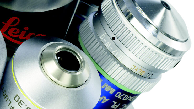
DM4 B & DM6 B
Microscopi verticali
Microscopi ottici
Prodotti
Home
Leica Microsystems
DM4 B & DM6 B Microscopi diritti
Risultati rapidi, flussi di lavoro personalizzati e informazioni approfondite
Leggi gli articoli più recenti
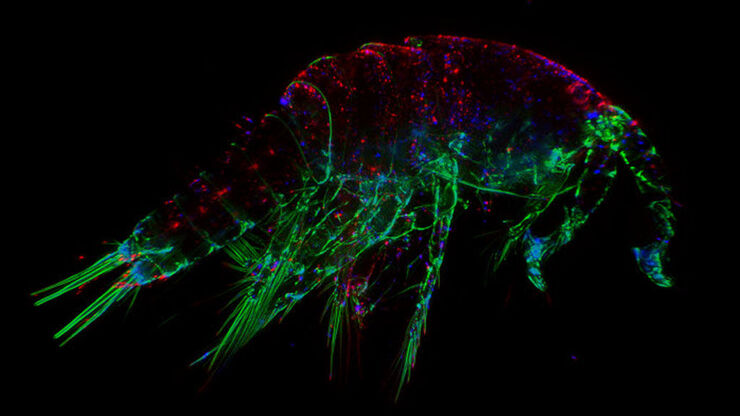
, Tropomyosin (cardiomyocytes, red) and GFP (primordial cardiac layer, green).")
A Guide to Fluorescence Microscopy
Fluorescence microscopy uses the ability of fluorophores, dyes, or fluorescent proteins to emit light of a specific wavelength after being excited with light of a shorter wavelength. Biomolecules can…

Factors to Consider When Selecting a Research Microscope
An optical microscope is often one of the central devices in a life-science research lab. It can be used for various applications which shed light on many scientific questions. Thereby the…

Improving Zebrafish-Embryo Screening with Fast, High-Contrast Imaging
Discover from this article how screening of transgenic zebrafish embryos is boosted with high-speed, high-contrast imaging using the DM6 B microscope, ensuring accurate targeting for developmental…

Epi-Illumination Fluorescence and Reflection-Contrast Microscopy
This article discusses the development of epi-illumination and reflection contrast for fluorescence microscopy concerning life-science applications. Much was done by the Ploem research group…

Differential Interference Contrast (DIC) Microscopy
This article demonstrates how differential interference contrast (DIC) can be actually better than brightfield illumination when using microscopy to image unstained biological specimens.
 cells taken with phase contrast.")
Phase Contrast and Microscopy
This article explains phase contrast, an optical microscopy technique, which reveals fine details of unstained, transparent specimens that are difficult to see with common brightfield illumination.
Immersion Objectives
How an immersion objective, which has a liquid medium between it and the specimen being observed, helps increase the numerical aperture and microscope resolution is explained in this article.

Microscopi per campo scuro
Il metodo di contrasto in campo oscuro sfrutta la diffrazione o la dispersione della luce da strutture di un campione biologico o da caratteristiche non uniformi nella struttura di un materiale.

Contrasto di fase
Grazie al microscopio ottico a contrasto di fase è possibile visualizzare le strutture di molti tipi di campioni biologici con maggior contrasto senza la necessità di utilizzare le colorazioni.

Studying Pulmonary Fibrosis
The results shown in this article demonstrate that fibrotic and non-fibrotic regions of collagen present in mouse lung tissue can be distinguished better with polarized light compared to brightfield.…

Virologia
L’oggetto della tua ricerca si concentra su infezioni e malattie virali? Scopri in che modo puoi approfondire l’aspetto virologico utilizzando le soluzioni di imaging e la preparazione del campione di…
The Fundamentals and History of Fluorescence and Quantum Dots
At some point in your research and science career, you will no doubt come across fluorescence microscopy. This ubiquitous technique has transformed the way in which microscopists can image, tag and…

Koehler Illumination: A Brief History and a Practical Set Up in Five Easy Steps
In this article, we will look at the history of the technique of Koehler Illumination in addition to how to adjust the components in five easy steps.
 illuminated with wide-band UV excitation. Note the tissue structure with blue autofluorescence. Right: Same tissue and same immunostaining with FITC label illuminated with epi-illumination using narrow")
Milestones in Incident Light Fluorescence Microscopy
Since the middle of the last century, fluorescence microscopy developed into a bio scientific tool with one of the biggest impacts on our understanding of life. Watching cells and proteins with the…

Video Tutorial: How to Align the Bulb of a Fluorescence Lamp Housing
The traditional light source for fluorescence excitation is a fluorescence lamp housing with mercury burner. A prerequisite for achieving bright and homogeneous excitation is the correct centering and…

Video Tutorial: How to Change the Bulb of a Fluorescence Lamp Housing
When applying fluorescence microscopy in biological applications, a lamp housing with mercury burner is the most common light source. This video tutorial shows how to change the bulb of a traditional…

Optical Contrast Methods
Optical contrast methods give the potential to easily examine living and colorless specimens. Different microscopic techniques aim to change phase shifts caused by the interaction of light with the…

Forensic Detection of Sperm from Sexual Assault Evidence
The impact of modern scientific methods on the analysis of crime scene evidence has dramatically changed many forensic sub-specialties. Arguably one of the most dramatic examples is the impact of…
Legga i nostri ultimi articoli
Microscopi per la ricerca Life Sciences
I microscopi per la ricerca Life Sciences di Leica Microsystems supportano le esigenze di imaging della comunità scientifica con la massima capacità innovativa e la competenza tecnica ai fini della…
Patologia clinica
Scoprite in che modo le soluzioni Leica per la microscopia per anatomia patologica aiutano i patologi clinici a diagnosticare infezioni e malattie da liquidi e dei tessuti corporei.
Soluzioni di microscopia per QA e QC dei dispositivi medicali
Per un'ispezione e un controllo di qualità efficienti e confortevoli dei dispositivi medicali: scoprite come i microscopi Leica ti possono aiutare a raggiungere questo obiettivo.
