
Specialità mediche
Specialità mediche
Esplora una raccolta completa di risorse scientifiche e cliniche su misura per gli operatori sanitari, tra cui approfondimenti tra pari, casi clinici e simposi. Progettata per neurochirurghi, oculisti e specialisti in chirurgia plastica e ricostruttiva, otorinolaringoiatria e odontoiatria. Questa raccolta evidenzia gli ultimi progressi nel campo della microscopia chirurgica. Scoprite come le tecnologie chirurgiche all'avanguardia, come la fluorescenza AR, la visualizzazione 3D e l'imaging OCT intraoperatorio, consentano di prendere decisioni sicure e di essere precisi in interventi chirurgici complessi.
What is a Field-of-View Scanner?
A field-of-view scanner is an assembly of galvanometric scanning mirrors used in single-point confocal microscopes that offer the correct optical recording of large field sizes. The field-of-view…
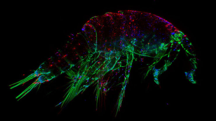
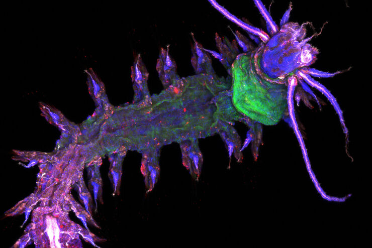
The Fundamentals and History of Fluorescence and Quantum Dots
At some point in your research and science career, you will no doubt come across fluorescence microscopy. This ubiquitous technique has transformed the way in which microscopists can image, tag and…

Eyepieces, Objectives and Optical Aberrations
This article covers the components of the eyepieces and how to adjust them correctly to suit your eyes.

Koehler Illumination: A Brief History and a Practical Set Up in Five Easy Steps
In this article, we will look at the history of the technique of Koehler Illumination in addition to how to adjust the components in five easy steps.

Collecting Light: The Importance of Numerical Aperture in Microscopy
Numerical aperture (abbreviated as ‘NA’) is an important consideration when trying to distinguish detail in a specimen viewed down the microscope. NA is a number without units and is related to the…

Introduction to Widefield Microscopy
This article gives an introduction to widefield microscopy, one of the most basic and commonly used microscopy techniques. It also shows the basic differences between widefield and confocal…
Optimization of the Interplay of Optical Components for Aberration Free Microscopy
Optical microscopes are used to magnify objects which are otherwise invisible for the human eye. For this purpose high quality optics is necessary to achieve appropriate resolution. However, besides…
 illuminated with wide-band UV excitation. Note the tissue structure with blue autofluorescence. Right: Same tissue and same immunostaining with FITC label illuminated with epi-illumination using narrow")
Milestones in Incident Light Fluorescence Microscopy
Since the middle of the last century, fluorescence microscopy developed into a bio scientific tool with one of the biggest impacts on our understanding of life. Watching cells and proteins with the…

Factors for Selecting Student Microscopes
If chosen carefully, educational microscopes will open windows to a cosmos of minute detail which delight young minds in schools and universities – and ideally keeps them fascinated enough to make…
