THUNDER Imaging Systems
THUNDER Imaging Systems
キーファクト Show subnavigation
THUNDER Imaging Systems
Leica Science Lab Show subnavigation
Read our latest articles about THUNDER Imaging Systems
The knowledge portal of Leica Microsystems offers scientific research and teaching material on the subjects of microscopy. The content is designed to support beginners, experienced practitioners and scientists alike in their everyday work and experiments.

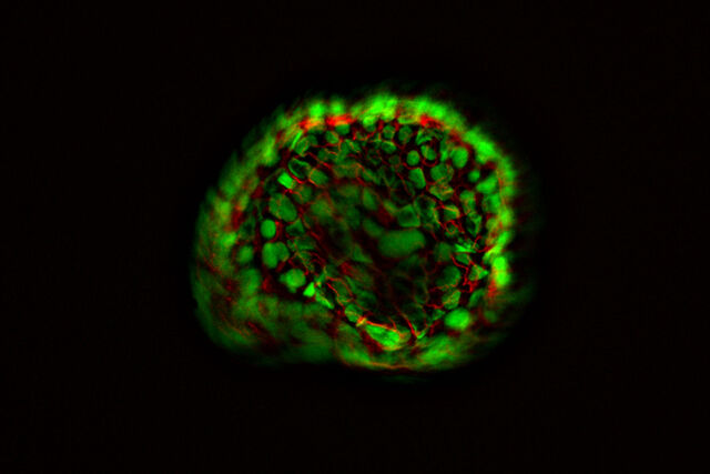
Unlocking the Secrets of Organoid Models in Biomedical Research
Designing the Future with Novel and Scalable Stem Cell Culture
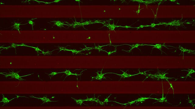
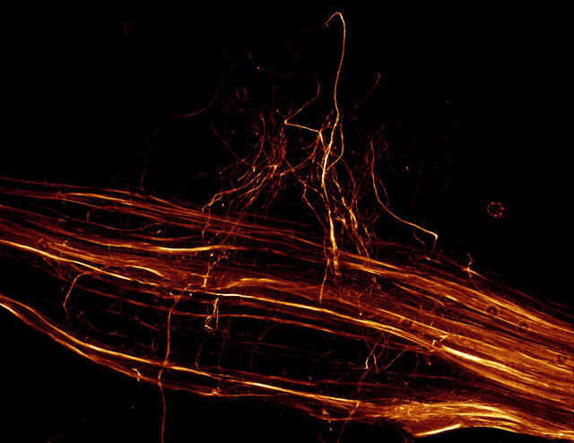
神経細胞移動の分子的秘密を解き明かす
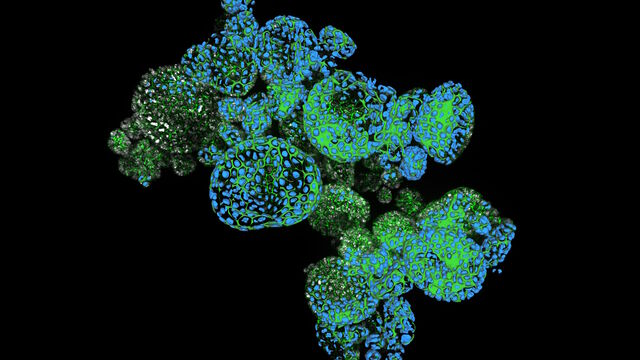
How Efficient is your 3D Organoid Imaging and Analysis Workflow?
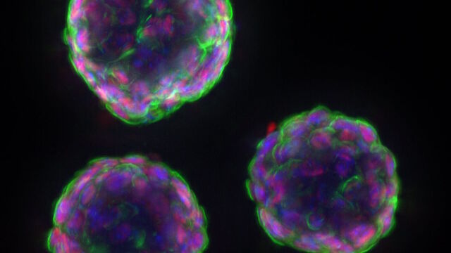
How to Get Deeper Insights into your Organoid and Spheroid Models
Imaging Organoid Models to Investigate Brain Health
機械受容性経路とシナプス経路の研究に顕微鏡がいかに役立つか
What are the Challenges in Neuroscience Microscopy?
The Role of Iron Metabolism in Cancer Progression
Going Beyond Deconvolution
Find Relevant Specimen Details from Overviews
Accurately Analyze Fluorescent Widefield Images
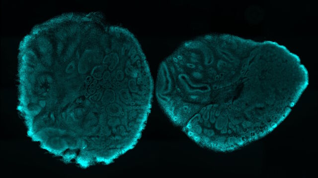
High-resolution 3D Imaging to Investigate Tissue Ageing
How to Improve Live Cell Imaging with Coral Life
Optimizing THUNDER Platform for High-Content Slide Scanning
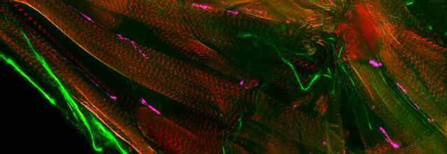
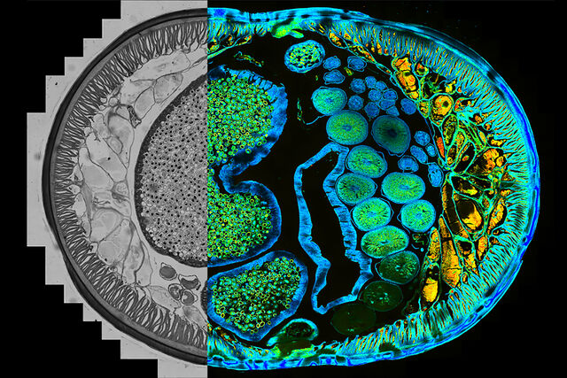
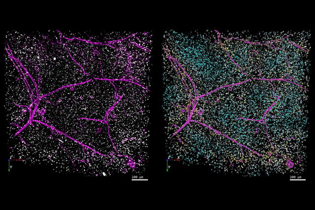
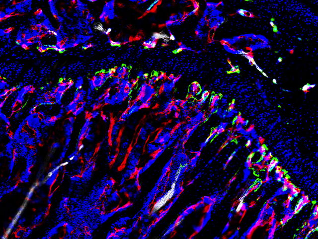
Physiology Image Gallery
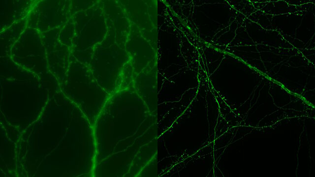
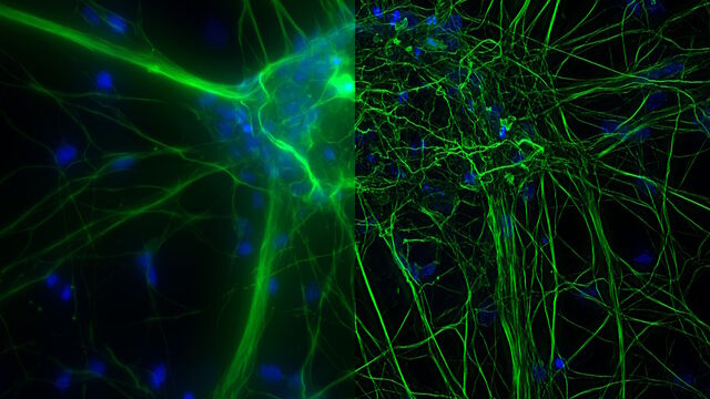
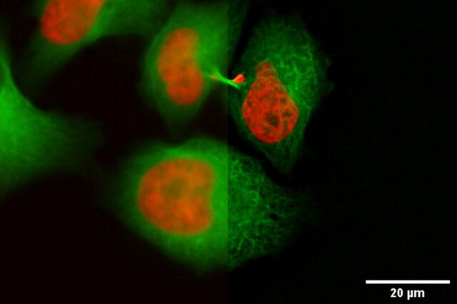
 従来の蛍光画像THUNDER画像
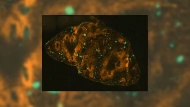
HeLa 細胞(標識;アクチン_Alexa Fluor 568 ファロイジン、核_YOYO 1 iodide)

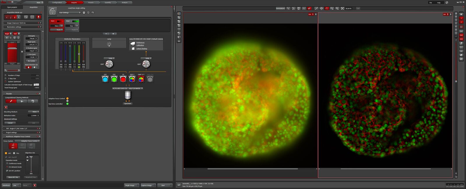
THUNDER の技術
THUNDER は、最新の Computational Clearing 法を用いて高解像度、高コントラストの画像を計算処理で提供するデジタルオプティクス技術です。Computational Clearing は、厚いサンプルでしばしば見られるフォーカスアウトしたボケ情報を除去します。これにより、z スタックでも深部の一平面画像でも優れた結果が得られます。
ライカの THUNDER テクノロジーは、ボケのない画像をリアルタイムに取得するため、関連する光学的パラメータをすべて考慮します。
テクノロジーノート
まだ疑問な点がありますか?THUNDERテクノロジーに関するより詳細な解説は、テクノロジーノートをご覧ください。
 従来の蛍光画像THUNDER画像
バッタの神経節、従来の蛍光画像(左)、THUNDER 像(右)。厚さ:110µm、データ量:376 MB。Computational Clearing による画像取得時間:3 秒
「大型サンプルでも 2 分以内に驚くほど鮮明な画像を提供する THUNDER はタイムラプス試験に有効です」
