
Industrial
Industrial
Mergulhe fundo em artigos detalhados e webinars com foco em inspeção eficiente, fluxos de trabalho otimizados e conforto ergonômico em contextos industriais e patológicos. Os tópicos abordados incluem controle de qualidade, análise de materiais, microscopia em patologia, entre muitos outros. Este é o lugar onde você obtém insights valiosos sobre o uso de tecnologias de ponta para melhorar a precisão e a eficiência dos processos de fabricação, bem como o diagnóstico e a pesquisa patológicos precisos.

Organismos Modelo na Pesquisa
Um organismo modelo é uma espécie usada pelos pesquisadores para estudar processos biológicos específicos. Eles têm características genéticas similares aos seres humanos e são usados tipicamente em…

Microscopy in Virology
The coronavirus SARS-CoV-2, causing the Covid-19 disease effects our world in all aspects. Research to find immunization and treatment methods, in other words to fight this virus, gained highest…

Pesquisa de câncer
O câncer é uma doença complexa e heterogênea causada por células com deficiência na regulação do crescimento. Mudanças genéticas e epigenéticas em uma ou em um grupo de células prejudicam o…
Digital Classroom Options
As teachers, you know your big challenge is to catch and keep the students’ attention and the best chance for this is by making the environment interactive. In the case of the Microscopy Classroom, we…
 images")
How To Create EDOF (Extended Depth of Focus) Images
Watch this video to see how you can rapidly record sharp optical microscope images of samples with a large height variation. This is done with the optional Extended Depth of Focus (EDOF) function of…
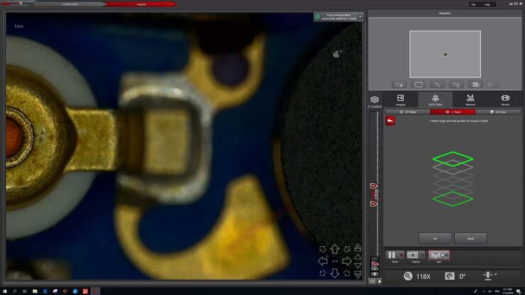
How to Make a Fast Z-stack
Save time for your 2D and 3D analysis. Watch this video to learn about the new user interface, LAS X.next, for the DVM6 digital microscope. The video demonstrates how to make a fast Z-Stack with a few…

Digital Microscopy in Earth Science
Classical polarized light (compound) microscopes can only be used for prepared samples, because the working distance they offer is insufficient for whole samples. This means that thicker and bigger…

Introduction to Mammalian Cell Culture
Mammalian cell culture is one of the basic pillars of life sciences. Without the ability to grow cells in the lab, the fast progress in disciplines like cell biology, immunology, or cancer research…

Introduction to Widefield Microscopy
This article gives an introduction to widefield microscopy, one of the most basic and commonly used microscopy techniques. It also shows the basic differences between widefield and confocal…
